
13 May - Muscoloskeletal
Program
1000-1015: Intro and recap from last week Stuart McNamara
1015-1115: Ankle and wrist #: Mr Jarrad Stevens (orthopaedic surgeon)
1115-1125: Coffee break – which beans today?
1125-1155: #NOF- Sophie
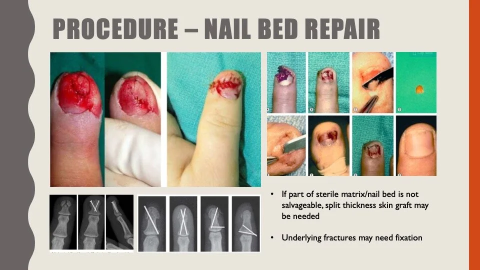
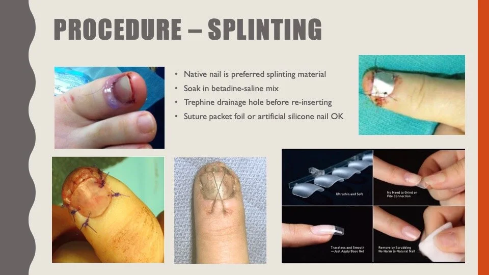
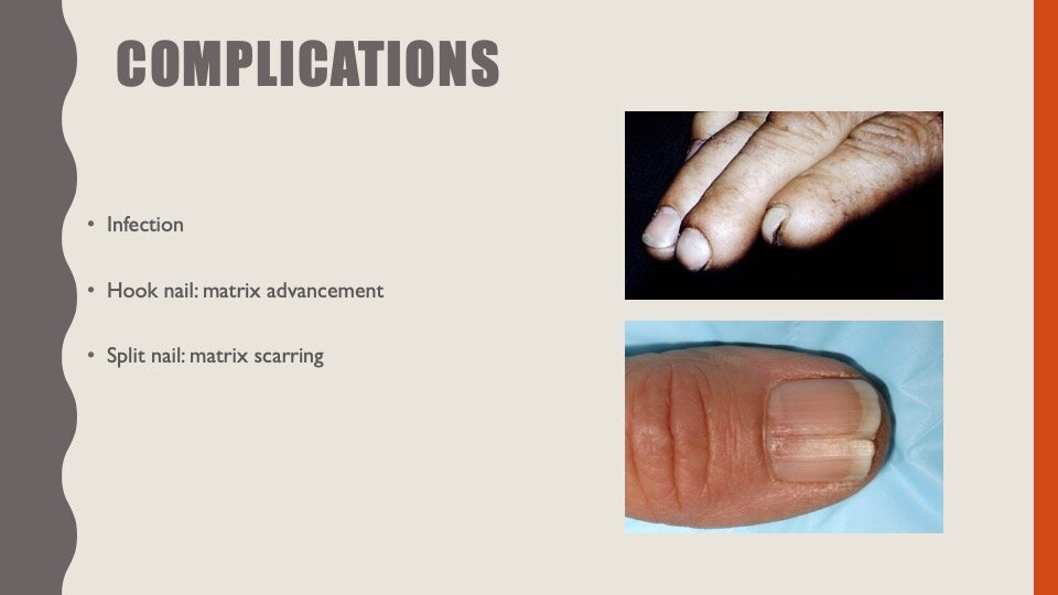
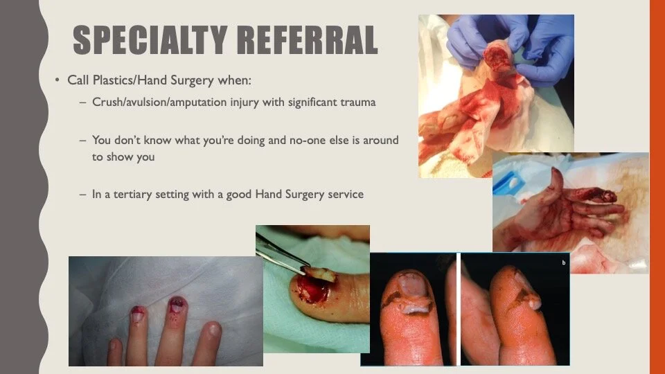
1155-1215: Nail bed injuries – Sallyanne
1215- 1250: ECG / Image / Gas of the week with Alex, Matt and Mark
1250-1300: 100 Burpee challenge – get into your active wear!!
1300-1400: M & M with Kirsten
1300 - 1500: OSCE Practice - Brendan
Key Learning Points
Distal Radius Fractures
St Vincent’s has a pathway for the management of distal radius fractures. This contains all the essential information on managing these patients. Click on the image to enlarge.
For further practical advice on the management of fractures, Jarrad highly recommends the book “The close treatment of common fractures” - John Charnley.
Key tip on reducing wrist fractures - reduce any impaction and then exaggerate the fracture to engage the periosteum.
All patients with distal radius fractures should be prescribed vitamin C for three months. Evidence suggests that this decreases the risk of chronic regional pain syndrome (CRPS). Given how debilitating this syndrome can be, the safety profile of vitamin C and the inability to predict which patients will go on to develop CRPS it is advised that we treat all patients. Prescribe 500mg daily. https://www.ncbi.nlm.nih.gov/pubmed/28274883
Fractured NOF
We see these injuries all the time. Remember that they almost always require a CXR and an IDC. These injuries convey a relatively higher mortality over the next year for the patient than you may think.
In the ED, we need to be experts in providing pain relief. This includes the use of regional analgesia. There is always some debate on the effectiveness of fascia iliac blocks vs femoral nerve blocks.
I recommend the following website to help learn how to do these blocks.
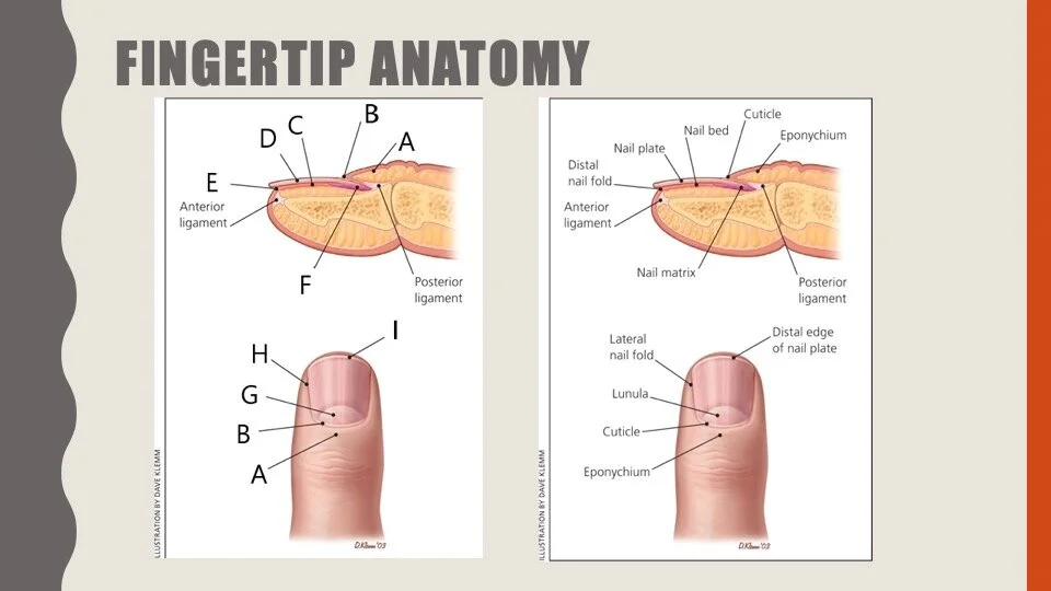
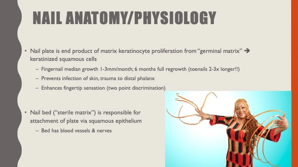
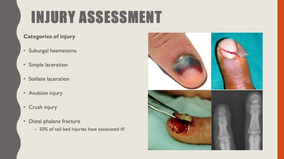
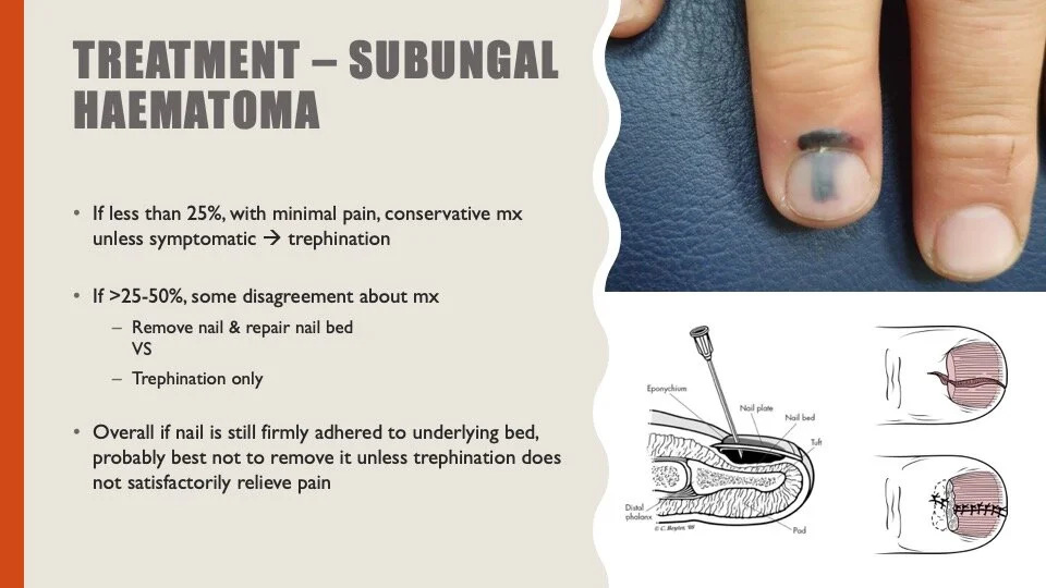
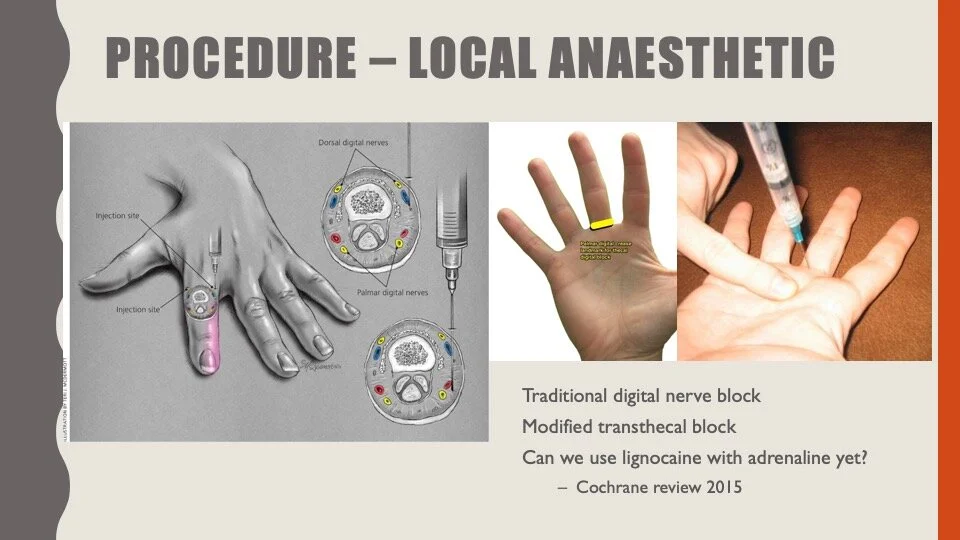
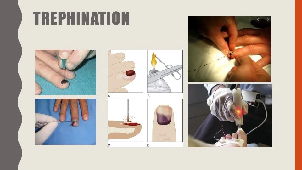
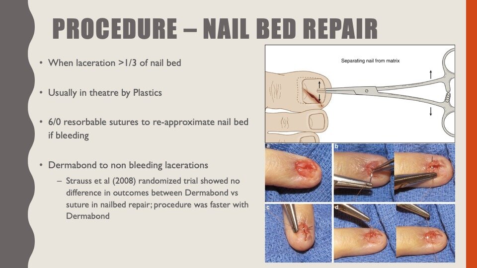
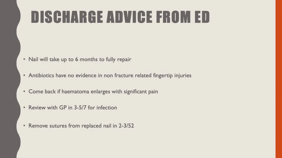
Nail-bed injuries
I have included the slides for this talk below.



ECG of the Week
Dino sent in this ECG from last week.
The patient presented with 2.5 hours of chest pain. He looked unwell and was in distress. Vital signs within normal limits.
He is 58 years old. Weight 75kg
This ECG shows ST depression in V2-V4. This ECG is suggestive of a possible isolated posterior STEMI and should prompt you to ask for an ECG with posterior leads. If those leads were normal then this ECG would be consistent with a NSTEMI with anterior ischaemia.
Most posterior STEMIs occur in conjunction with a inferior STEMI and therefore less clinically significant as the patient requires reperfusion therapy anyway. However, it is estimated hat 5-10% of all STEMIs are isolated to the posterior region and will present with ECGs like this. They are commonly missed.
When diagnosed, there is a clear indication for cath lab activation for patients with ST elevation in the posterior leads.
This is posterior ECG of this patient. Click to enlarge. The patient went to Cath lab and had a successful PCI.
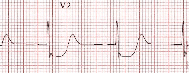
On the normal 12 lead, a posterior STEMI is suggested by horizontal ST depression in the anterior leads with an upright T wave.
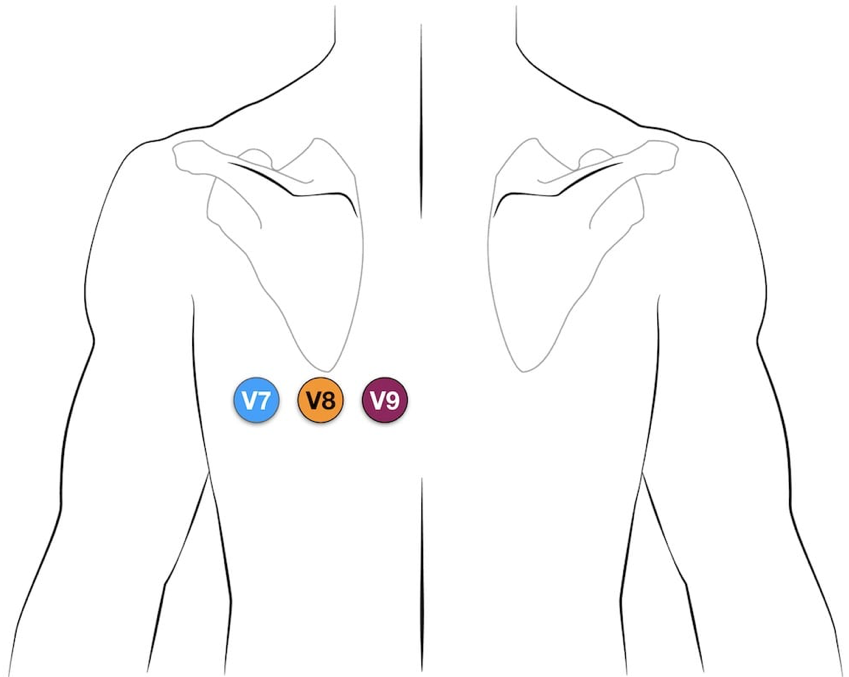
Posterior leads are easy. Take V4,V5,V6 off the patient and place on the back with V8 at the tip of the scapula
Interesting Blood Gas
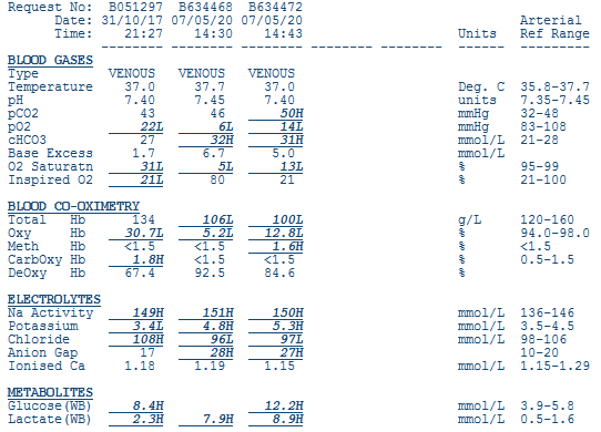
72 yo female presents via ambulance with dark coloured vomiting and abdominal discomfort.
Symptoms began in the previous 6 hours.
PMHx: Bilateral hip surgery, immobile requiring hoist transfers post op. Ischaemic heart disease.
No regular medications
HR 150, BP 85/65, SpO2 88% RA, RR 24, Temp 38.5, GCS 15
Looks unwell
Abdomen is generally tender to palpation.
Look at the VBG on the far right from 14:43
What is going on?
(Albumin 26)
Here is Mark’s interpretation of this gas.
F-S approach:
Overall metabolic alkalosis as the BE is positive. pH normal so is there a compensatory respiratory acidosis? Can’t tell as a venous gas.
SID effect: 150 + 5 – 97 = 58.
Should be 42 therefore 16 too few chloride i.e a hypochloraemic alkalosis. This should give a BE of + 16.
Albumin effect: alb 26, should be 42, there is a hypoalbuminaemic alkalosis
Alb effect: (42-26)/4 = 4. This should give a BE + 4.
Lactate effect = BE -8
Total measured effects = 16 + 4 – 8 = +12.
Actual BE = 5 therefore -7 “unexplained” = metabolic acidosis likely ketones.
Other considerations:
Na 150 = high
- likely represents free water deficit
- (150-140)/140 x 0.6 x weight (?70kg)
- = 3 litres
Putting it together:
Hypochloraemic alkalosis from vomiting, element contraction alkalosis too. Can I use N/Saline?
Lactic acidosis: systemic? (from hypovolaemia/hypoperfusion possibly aggravated by poor CO given PHx cardiac disease) versus regional? (ischaemic gut given abdo pain, known atherosclerotic disease)
Hypoalbuminaemia: chronic? (nutritional, CLD) vs subacute? (catabolic state after recent surgery, acute consumption due to sepsis etc)
Ketoacidosis – due to dehydration +/- starvation given abdo pathology
Resp acidosis?? Due to gut distention/splinting +/- aspiration etc
Old school approach:
Bicarb high, there is a primary metabolic alkalosis. pH normal; also resp acidosis??
Anion gap Na – Cl – HCO3 = 150-97-31 = 22. Normal AG is 12. There is a RAGMA of 10. (This approach UNDERESTIMATES extent of acidosis because it incorporates, but does not acknowledge, the small hypoalbuminaemic ALKALOSIS)
Delta gap: Delta AG = 22-12 = 10. Delta Bicarb = 26 – 31 = -5.
Therefore delta gap 15. Bicarb should drop by 1 for every 1 AG is increased if only have RAGMA; expect bicarb to drop by 10. Actual bicarb INCREASED by 5; there is a +15 ‘effect’ unexplained = metabolic alkalosis. This represents hypochloraemia.
Or:
Delta ratio = delta gap/bicarb gap = 10/-5 = -2
Figure that one out!!! Delta ratios suck
(Alex’s Peer review comment here - In this case the delta ratio is not helpful. Usually, it would only be suggested to do a delta ratio if there is a primary RAGMA)
Remember:
1. Delta gap/ratio is just a convoluted way of seeing if there is a chloride derangement if albumin is not provided.
2. If you do not adjust AG for hypoalbuminaemia, delta gap overestimates Cl problem if albumin is low.
3. A NAGMA = hyperchloraemic acidosis. Call it what it is!!!
4. Just calculate SID!!!
If this was an ARTERIAL gas, would need to calculate expected respiratory compensation
Expected pCO2 = 0.9 x HCO3 + 9 (+/-2)
Images of the Week
The images for this week come from the same 72 yo female as in the blood gas above. Click on the image to enlarge.
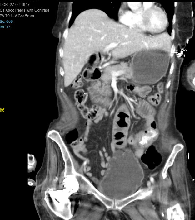
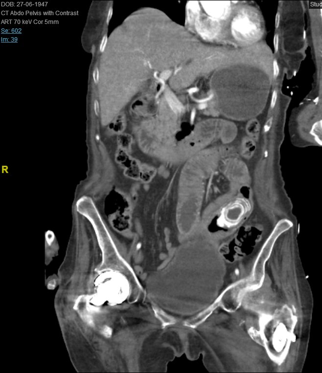
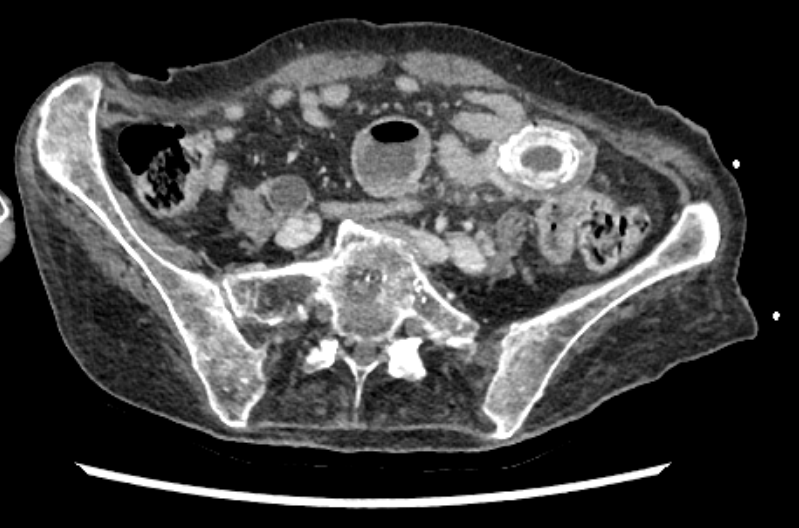
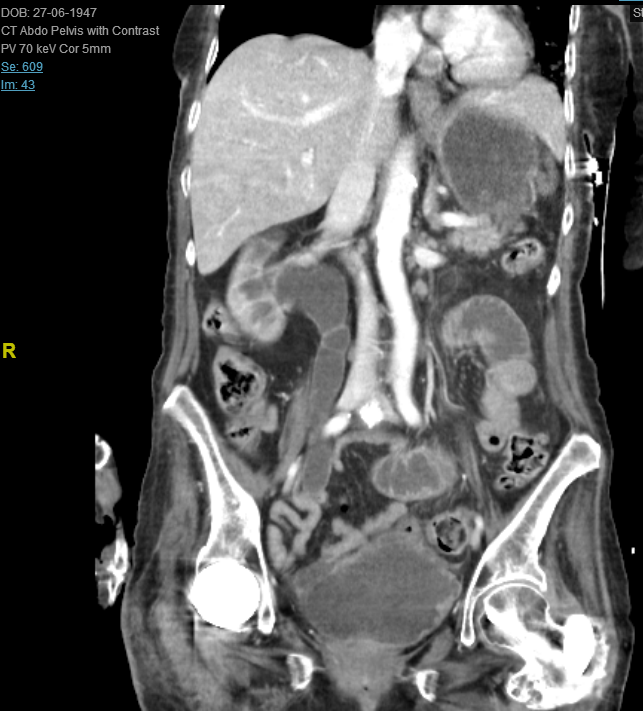
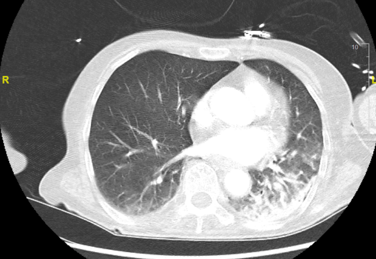
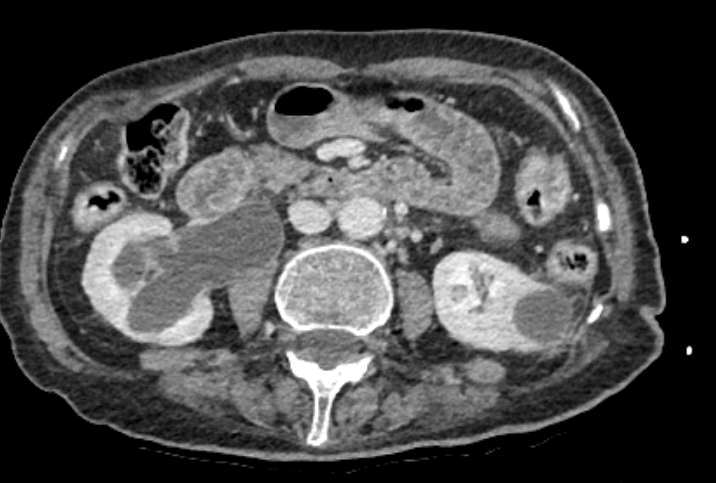
This CT shows a number of pathologies. These include:
3.5cm gallstone within the mid jejunum
Pneumobilia
Small bowel obstruction: dilated loops of small bowel, thickened bowel wall, air fluid levels
Right hydroureter and hydronephrosis. Left renal cyst
Left lower lobe consolidation
Matt discussed a fantastic approach for the emergency doctor looking at a CT. He essentially described looking for the things that will kill the patient first.
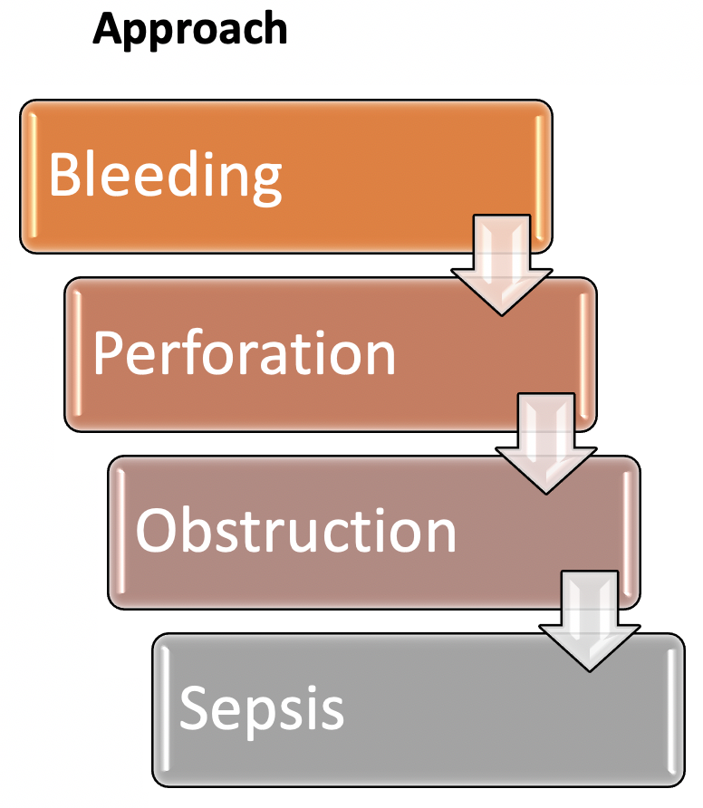
He suggests a 3 minute CTAP screen as described below.
3min CTAP Screen
Aorta down (axial plane):
2cm, no dilation or false lumen
Free fluid (axial plane):
Perihepatic
Morrisons pouch
Splenorenal angle
Paracolic gutters
Pouch of Douglas
Perforation (axial plane- bone window):
Uncontained free gas
Obstruction/ bowel pathology (axial plane):
3cm small bowel
6cm large bowel
9cm caecum
No fat stranding
Not thick walled
No gas in the wall
IA sepsis (axial & coronal)
Organs: lung, liver, gallbladder, spleen, kidneys, pancreas, bladder
Relook coronal