
Interpreting the Blood Gas
Alex Handrinos
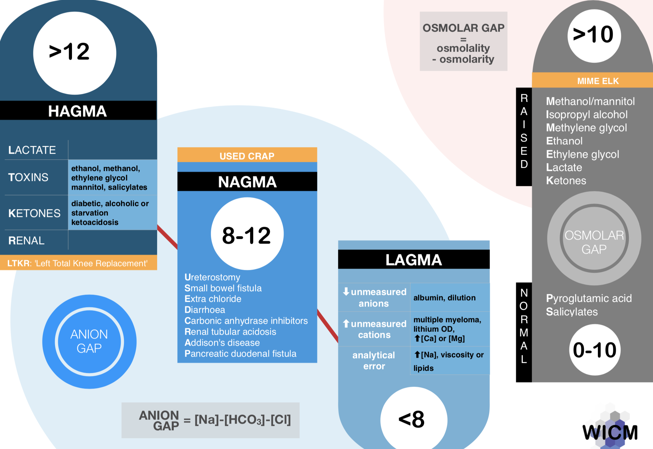
There are a number ways to interpret a blood gas. Each have their own advantages and disadvantages. Here I will explain the method that I used for the ACEM fellowship exam. It is a more ‘traditional’ method of interpretation using a bicarbonate approach and was developed largely from an approach published in Lawton’s Own the ABG book as well as this graphic made from Wellington ICU.
The approach is based on trying to extract as much information from the blood gas as possible (in order to answer the fellowship questions). The following general steps are involved:
Look at the acid base status
Look at the oxygenation (arterial samples only)
Look at the electrolytes and other values
Clinical interpretation
I have included a worked example at the end.

Step 1 - Acid Base Status
Identify the primary disturbance
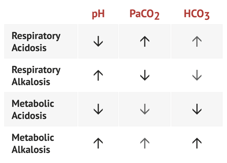
Comment on the pH. Is the patient academic or alkalaemic
Comment on the CO2. Is there hypercarbia or hypocarbia
Comment on the HCO3-. Is it high or low
Decide on the major disturbance. This will always be in the same direction as the pH
For example if the patient is acidaemic (pH < 7.35) and the bicarbonate is low the primary problem is a metabolic acidosis
If the patient is acidaemic (pH < 7.35) and the pCO2 is high the primary problem is a respiratory acidosis
Identify any additional disturbances by calculating expected values
If the primary disturbance is metabolic then calculate the expected pCO2.
When the primary disturbance is a metabolic acidosis then use Winters’ formula:
Expected pCO2 = 1.5 x HCO3- + 8
When the primary disturbance is a metabolic alkalosis:
Expected pCO2 = 0.7 x HCO3 + 20
Interpretation: If the expected pCO2 is lower than the measured pCO2 it suggests a concurrent respiratory acidosis (even if the measured pCO2 is otherwise within normal range as the patient has not compensated as expected for the metabolic disturbance). In contrast, if the expected pCO2 is higher than the measured CO2 it suggests a concurrent respiratory alkalosis.
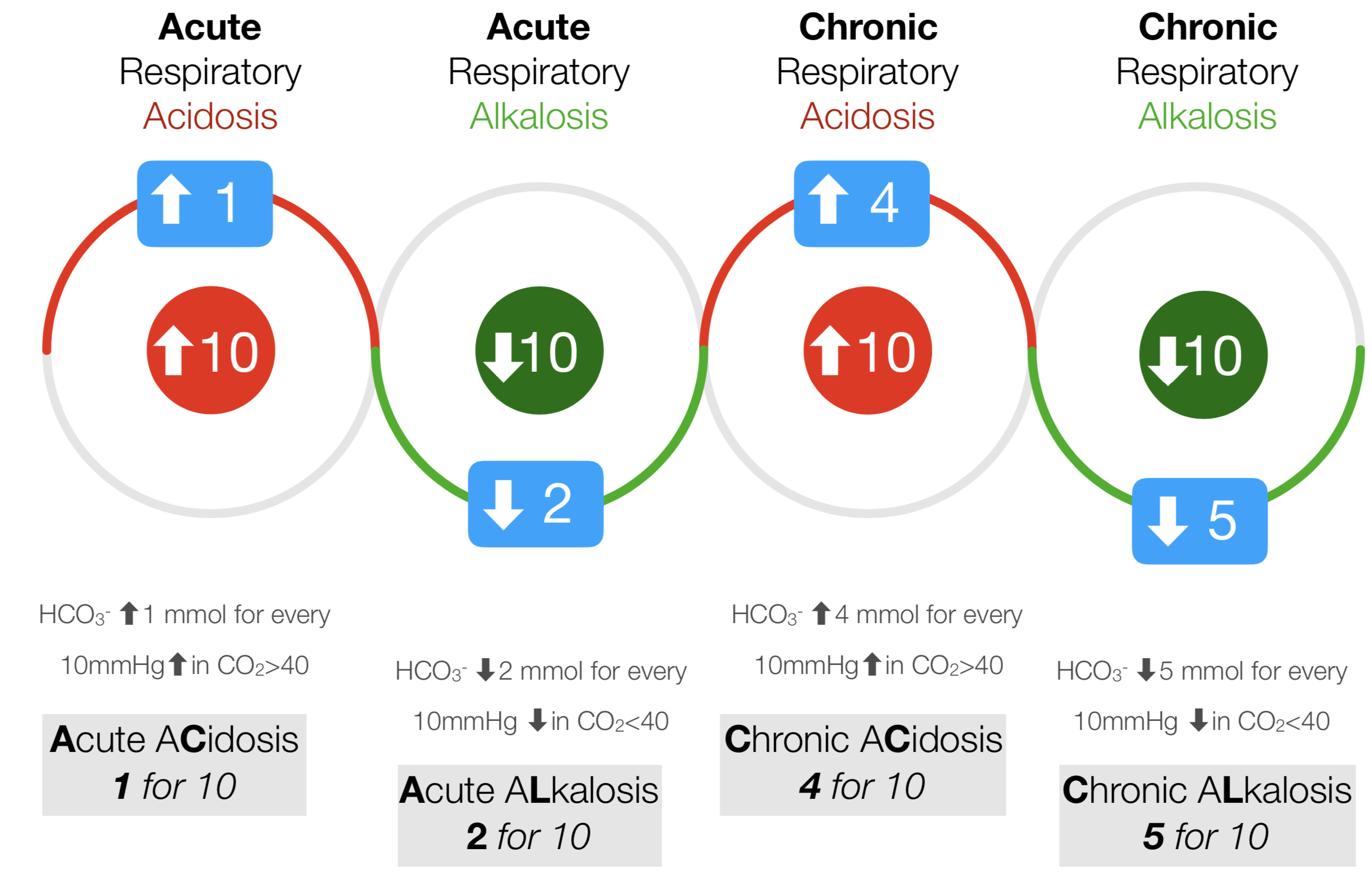
If the primary disturbance is a respiratory acidosis or alkalosis then calculate the expected HCO3-. The 1:2:4:5 rule can be used here
Acute respiratory acidosis: HCO3- increased by 1mmol for every 10mmHg in increase in CO2 above 40
Acute respiratory alkalosis: HCO3- decreased by 2mmol for every 10mmHg in decrease in CO2 below 40
Chronic respiratory acidosis: HCO3- increased by 4mmol for every 10mmHg in increase in CO2 above 40
Chronic respiratory alkalosis: HCO3- decreased by 5mmol for every 10mmHg in decrease in CO2 below 40
Arbitrarily, 24mmol/L is said to be the normal baseline HCO3- for the purpose of calculation.
Interpretation: If the expected HCO3 is lower that the measured HCO3- it suggests a concurrent metabolic alkalosis. If the expected HOC3- is higher than the measured HCO3 it suggests a concurrent metabolic acidosis.
If there are two disturbances occurring in the same direction (i.e. a primary metabolic acidosis and respiratory acidosis - think pH 6.9, pCO2 80 and bicarb 12) you can correct in both directions to prove to yourself that multiple processes are occuring.
Metabolic Acidosis
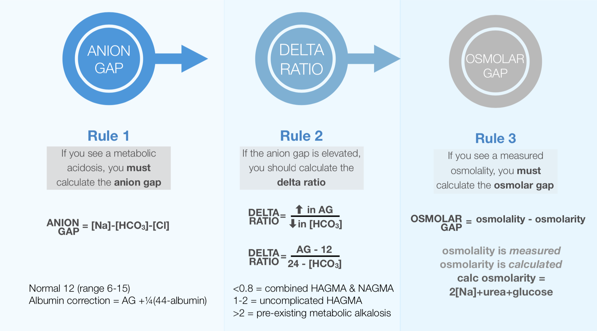
If a metabolic acidosis is detected then proceed with a calculation for anion gap (normal = 12) and if raised, a delta ratio.
Anion Gap = Sodium - Chloride - Bicarbonate
If the Anion Gap is > 12 calculate a delta ratio to evaluate a concurrent of either normal anion gap metabolic acidosis (NAGMA) or a metabolic alkalosis
Delta Ratio = (AG-12)/(24 - HCO3-)
If an osmolality is provided (in an exam setting) then make sure to calculate the osmolar gap (more on this later).
What about the Fencl-Stewart approach? This an alternative to using anion gap and delta ratio and can be read about here.
Step 2 - Oxygenation
Look at the O2 Sats (sO2) - is this definitely an arterial sample?
Look at the pO2 - is the patient hypoxaemic?
In the context of the patient, is the methaemoglobin or carboxyhemoglobin level important?
A-a Gradient
This is only applicable to arterial blood samples and only really relevant if the patient is hypoxic. There are number of different ways to calculate the A-a gradient. Ultimately it doesn’t matter as the exact number is not as important as knowing whether the A-a gradient is normal or raised. I find the simplest method below.
A-a Gradient = PAO2 (alveolar - calculated) - PaO2 (arterial - measured)
= (760-vapour pressure) x FiO2 - 1.25x PaCO2) - PaO2
The most challenging part is trying to work out the first part of the equation. If the patient is on room air (FIO2 21%) and presumed to be at sea-level then (760-vapour pressure)) x FiO2 = 150mmHg. The equation would then become Aa gradient = 150 - 1.25 x PaCO2 - PaO2
If the patient is on FiO2 40% the value becomes 300mmHg
If the patient is on FiO2 80% the value becomes 600mmHg
If the patient is on FiO2 100% the value is 713mmHg (700 acceptable for the purpose of calculation)
This estimation allows much simpler calculations that can be done rapidly in an exam setting.
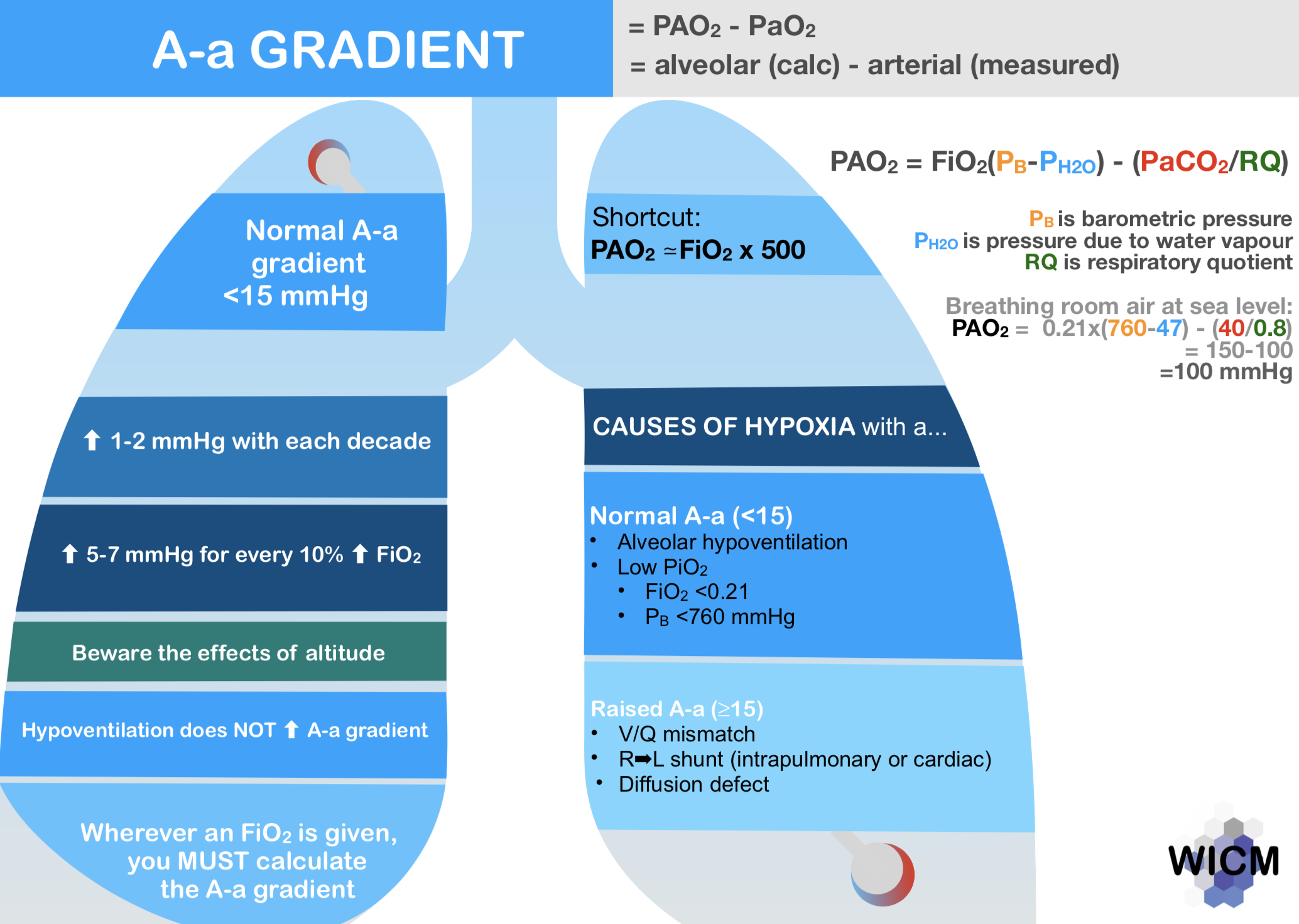
Interpretation: An A-a gradient > 15 suggests a V/Q mismatch. Examples include:
PE
APO
ARDS
LRTI
Other respiratory membrane disease.
Note 1: Hypoventilation does not cause a raised A-a gradient
Note 2: Expected Aa gradient = age/4 +4. Does this equation look familiar? It is the same as the uncuffed ETT tube size for paediatric patients.
Identifying a raised Aa gradient can assist in the diagnosis for hypoxia, although in my opinion this is largely academic and other than in exams I haven’t found this calculation particularly important in changing my emergency department management.
Step 3 - Electrolytes and other values
Look for and comment on any electrolyte disturbance. Remember to:
Correct Na for glucose
Correct K for pH
Calculate osmolality and osmolar gap
Identifying electrolyte disturbances and correcting appropriately can help direct where replacement/removal of electrolytes is required.
Sodium
There are many different methods for correcting the sodium in cases of hyperglycaemia. The easiest to remember is:
Corrected Na = Na + (Glucose-5)/3
Potassium
Potassium homeostasis is complicated and varies with the type and mechanism of academia or alkalaemia. Practically, and for the purpose of an exam for each fall of 0.1 in pH below 7.4, the potassium should rise by 0.5mmol above 5.0mmol/L. Conversely, for each rise of 0.1 in pH above 7.4, the potassium should drop by 0.5mmol below 5.0mmol/L
For example, if the pH is 6.9 and the measured potassium is 5.0mmol/L it would be reasonable to say: the expected potassium for this pH would be 7.5 therefore the patient has a relative hypokalaemia
Another way of looking at this is to say this is that the corrected potassium for this patient is 2.5mmol/L. Where the calculated outcome is the correction to the measured pH rather than the potassium that would be expected.
Not being aware of both methods can be confusing. It doesn’t matter which you use, just stick with it and know what it means
Osmolality
If an osmolality is given, it is essential to calculate the osmolar gap. Remember that:
OsmolaLity is measured in the Lab
OsmolaRity is calculated
Osmolar Gap = Osmolality - osmolarity
Osmolarity = 2 x Na + Urea + Glucose
Interpretation: Osmolar gap > 10 is considered raised. Causes of raised osmolar gap include toxic alcohols, mannitol use, lactate and ketones
Step 4 - Clinical interpretation
It is all well and good to be able to churn out the calculations described above. But why are we doing this? What information can I gather from the above information to help me with diagnosis and guidance of patient care?
Identifying the presence of primary and secondary disturbances can help lead to a list of differential diagnoses as below.
Causes of HAGMA
Mudpiles, Goldmark, Dumsale is out. Too complicated and when was the last time you saw an isoniazid overdose?
I use the acronym LTKR - left total knee replacement, which will cover just about any relevant cause of a raised lactate.
L - Lactate
T - Toxins
K - Ketones
R - Renal
Lactate is by far the most common cause of a high anion gap metabolic acidosis. It is important to know that there are many causes of a raised lactate and this classification (Cohen & Woods) is important in itself. Common examples listed below:
Type A - Inadequate oxygen delivery
Anaerobic muscle activity (convulsions, post wrestling)
Tissues hypoperfusion (shock, cardiac arrest, mesenteric ischaemia)
Reduced tissue oxygen delivery (hypoxaemia, anaemia) or utilisation (CO poisoning)
Type B - No evidence of inadequate tissue oxygen delivery
Type B1 - Associated with underlying disease
LUKE: Leukaemia, lymphoma
TIPS: Thiamine deficiency, infection, pancreatitis, short bowel syndrome
FAILURES: hepatic, renal, diabetic failures
Type B2 - Associated with drugs and toxins
Long, long list: big ones include cyanide, salicylates, paracetamol, metformin
Type B3 - Associated with inborn errors of metabolism
Raised ketones are due to one of three reasons
Diabetic emergency i.e DKA
Alcohol ketoacidosis
Starvation Ketoacidosis
Toxins are covered above in causes of a raised lactate.
Renal failure and acid base is complex with multiple mechanisms that can explain an acidosis. Uraemic renal failure can also lead to a HAGMA due to the failure to excrete acid anions. Note that “renal failure” as an entity can also cause a NAGMA due to renal tubular acidosis where there is an impaired ability to acidify the urine with net acid retention and hyperchloraemia.
Causes of NAGMA
There are many acronyms out there. I use ABCD as my memory aid.
A - Adrenal crisis/Addisons
B - Bicarbonate loss (most common) - excessive diarrhoea, renal tubular acidosis, fistulas (e.g. pancreaticoduodenal)
C - Chloride - excessive NaCL administration
D - Drugs - acetazolamide, spironolactone
Below is the Wellington ICU approach to interpreting a metabolic acidosis which differs slightly from my own.
Causes of metabolic alkalosis
Again, many memory aids available. I use GROE
G - GI Acid Loss - Vomiting (most common), diarrhoea, Ileostomy
R - Renal Acid loss - Diuretics, Bartter/Gitelman syndrome, milk-alkali syndrome
O - Overdose of base - antacids, laxatives, iatrogenic HCO3 administration
E - Endocrinopathy - Steroid excess, Cushing syndrome
Causes of respiratory acidosis
Generally speaking there are only a few causes of a respiratory acidosis including hypoventilation, increased CO2 production such as in malignant hyperthermia and increased inspired CO2 (rebreathing). The challenge is determining why there is hypoventilation - ? toxidrome, ? V/Q mismatch
Causes of respiratory alkalosis
This will always be caused by hyperventilation. Again, why is the patient hyperventilating. ? aspirin overdose, ? panic attack, ? ventilator respiratory rate too high.
Example
A 40 yo male multi-trauma patient attends the hospital after a high speed MVA. The ambulance have given him several litres of crystalloid en route to the hospital. He has obvious chest injuries.
Vitals
HR 110, BP 95/75, RR 34, SpO2 92% on 0.4 FIO2, GCS 7
Consider the following arterial blood gas:
FiO2. 0.4
pH 7.09
pO2 76 mmHg
pCO2 65mmHg
HCO3- 14 mol/L
BE -8
O2 Sats 92%
Na+ 135 mmol/L
K+ 4.2 mmol/L
Cl- 103 mmol/L
Glucose 6.2 mmol/L
Lactate 4.1mmol/L
Hb 82g/L
Step 1 - Acid Base
The patient has a severe acidaemia (pH 7.09) with a high pCO2 and low bicarbonate
Calculations
Expected PCO2 for HCO3 of 14
= 1.5 x 14 + 8
= 29
Therefore as the measured pCO2 is 65 there must be a concurrent respiratory acidosis
Expected HCO3 for pCO2 of 65
if Acute: 24 + (2.5 x 1) = 29mmHg
if Chronic: 24 + (2.5 x4) = 34mmHg
Therefore as the measured HCO3 is only 18 there must also be a metabolic acidosis
The primary disturbances are both a respiratory acidosis and metabolic acidosis.
As there is a metabolic acidosis present it is now important to check the anion gap and delta ratio
Anion Gap = Na - Cl - HCO3
= 135 - 103 - 14
= 18
Delta Ratio = (AG - 12)/ (24 - HCO3)
= (18-12)/(24-14)
= 6/10
= 0.6
This implies both a HAGMA and NAGMA
Therefore our overall interpretation of the acid-base status of this blood gas is of a triple disturbance:
Respiratory acidosis
HAGMA
NAGMA
Step 2 - Oxygenation
This patient is hypoxic despite oxygen therapy. An A-a gradient can be calculated
A-a Gradient = PAO2 - PaO2
= (760-vapour pressure) x FiO2 - 1.25x PaCO2) - PaO2
= 300 - (1.25 x 65) - 76
= 134
Therefore this patient has a raised Aa Gradient
Step 3 - Electrolytes and other values
Expected potassium for pH 7.09 = 5 + 3 x 0.5 = 6.5mmol/L
The actual potassium is 4.2 suggesting a relative hypokalaemia
Also noted in the blood gas is a hyperlactataemia and a severe anaemia
Step 4 - Clinical interpretation
This patient is sick!
The arterial blood gas shows a triple disturbance of a respiratory acidosis, HAGMA and NAGMA.
The HAGMA is likely explained by the hyperlactaemia. Probably Type A due to poor tissue perfusion from hypovolaemic shock.
The NAGMA could be due to excessive crystalloid administration or possible renal failure.
There is a respiratory acidosis with a raised A-a gradient suggesting VQ mismatch which in this context could be due to pulmonary contusion, haemo/pneumothorax, ARDS/APO, fat embolism or PE.
Now that you have spent fifteen minutes interpreting this patient’s blood gas you probably should go back and resuscitate the patient!