
6 May 2020 - Geriatrics, Palliative Care
Program
10-1015hr – Welcome and recap of last week – Dale ‘stand in’ Jacqueline
1015-1100hr – How to manage your palliative patient – Hope
1100-1110hr – coffee break
1110-1140hr – Ouch – Falls in the Elderly – Marrillo
1140-1200hr – 10 min Tox Case – Khalid
1200-1215hr – ECG of the Week – Alex
1215-1230hr – *New* Xray of the week - Matt
1230-1245hr – Gas of the week – Mark
1245-1300hr –quick lunch break
1300-1345ish hr – SAQ questions – Jacqueline/Sarah
1300-1500hr – Fellowship OSCE practice – Brendan, venue TBA
Key Learning Points
Palliative Care
Hope discussed the emergency management of common palliative symptoms. Here are some of the key learning points:
Pain - the PRN dose of opioid should be 1/6 of the total 24 hour opiate dose
Hydromorphone is a useful opioid in those with poor renal failure (eGFR < 40)
Use eviQ opioid calculator to help convert doses between different opioids and routes of administration - https://www.eviq.org.au/clinical-resources/eviq-calculators/3201-opioid-conversion-calculator
Subcutaneous doses of medications take 20-30 minutes to take effect
Syringe drivers do not work immediately - they will need PRN medications while taking effect (up to 4 hours)
Use haloperidol (0.5-1mg PO/SC/IM/IV) for nausea in palliative care - ondansetron will constipate!
Opiates are first line for treatment of dyspnoea, in small doses. I.e. ordine 1-2.5mg PRN
Always consider hypercalcaemia as a cause of altered conscious state. It can cause a whole range of different symptoms.
Treatment of hypercalcaemia in the ED is essentially just fluids - the rest of the treatment will happen on the wards
Always consider urinary retention in the palliative patient - get a bladder scan!
Falls in the elderly
1 year mortality after a fall is 15%
1 year mortality after hip fracture is 22%
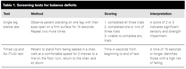
Be cautious when diagnosing a “mechanical fall”. Could this be a seizure, syncope, presyncope?
Several bedside tests can help to objectively assess the falls risk
Prevention of falls is so important. Encourage appropriate exercise, think about bone health, refer to falls clinic.
Tox Case
Khalid presented a case of a patient with verapamil and desvenlafaxine overdose
Verapamil
Resuscitate: haemodynamic support as below
Risk assessment: Ingestion 2-3x usual dose can lead to serious toxicity, 1-2 tablets in a child can kill, > 10 tablets in an adult is life threatening. Expect haemodynamic collapse
Supportive: As required
Investigations: ECG!
Decontamination: Charcoal up to 6 hours, longer if modified release
Enhanced elimination: Whole bowel irrigation possible in discussion with tox
Antidote: Calcium! 30mls Ca gluconate IV over 5 minutes. Repeat x 3 over the 1st hour. Infusions may be needed
Atropine for bradycardia
HIET: 50mls of 50% glucose followed by 1 unit/kg actrapid IV. Continue with 1unit/kg/hr infusion with ongoing glucose.
Inotropes
ECMO
methylene blue/lipid emulsion may be indicated in refractory cases
Disposition: Depends on dose. Prolonged observation as slow release
Desvenlafaxine
Resuscitate: treat seizures with diazepam, inotropic support if needed
Risk assessment: Seizures (all ingestions > 5g), seretonin toxicity, left ventricular dysfunction (if > 8g)
Supportive: As required
Investigations: Nil specific
Decontamination: Charcoal up to 4hours
Enhanced elimination: Whole bowel irrigation possible in discussion with tox
Antidote: none
Disposition: Depends on dose. Prolonged observation as slow release
ECG of the Week
90 yo female BIBA after being found on the floor.
This ECG is consistent with hypothermia. HR ~30, sinus bradycardia, shiver artifact, Osborne waves and a long QT
Other features of hypothermia that can be seen on ECGs include a slow AF, junctional bradycardia. Everything can be slowed - long PR, QRS, QT.
Osborne waves are small positive deflections at the J point. They are most prominent in praecordial leads. In hypothermia, the Osborne waves increase in size with decreasing temperature. Other causes of this notching include:
Hypothermia
Benign early repolarisation
Hypercalcaemia
Brugada
Neurological insult such as SAH
Image of the Week
Matt is going to start facilitating a radiology image of the week. Each week, have a look and have a go answering the questions anonymously below.
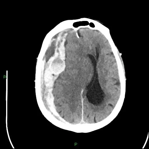
76 yo male BIBA from home alone after being found on the floor by his neighbours. PMHx: HT & AF on warfarin.
OE: HR 50, BP 186/120, GCS 13 on arrival which dropped to GCS 7 in CT
https://radiopaedia.org/cases/acute-subdural-haemorrhage-on-warfarin-2
Describe the key findings on the CT
1) Right subdural haematoma (mixed density blood, cystic appearance in places)
- Looks like acute-on-chronic but can be a hyper-acute bleed or effect of anticoagulation
- Convex, diffuse, & crosses suture lines
2) Significant midline shift
3) Effacement ipsilateral lateral ventricle, dilation of the contralateral lateral ventricle
4) Subfalcine herniation
Dilation of the contralateral ventricle
- Implies obstructive hydrocephalus from mass effect
- Requires urgent neurosurgical intervention
- The patient can rapidly deteriorate (as in this case)
Subfalcine herniation
- Implies critical raised intracranial pressure
- Another indication for urgent neurosurgical intervention
- Is an indication for ICP lowering treatment (mannitol, hypertonic saline, intubation with ongoing deep sedation & paralysis, and low normal PaCO2)
What are the risk factors for this pathology?
1) Cerebral atrophy: advanced age or chronic alcohol abuse
2) Anticoagulation
3) Chronic liver / kidney disease
4) Frequent falls
Interesting Blood Gas
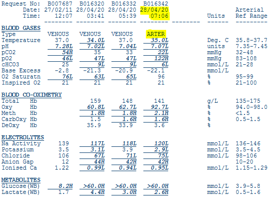
66 yo male presented with 4/7 of cough, malaise and abdominal pain. ATS 3 in COVID zone
PMHx: T2DM, HTN
Look at the arterial gas on the far right. The gas will be discussed in teaching this week.
The albumin was 38
Look at both blog posts on gas interpretation to assist if needed.
Primary Acid/Base
There is a severe acidaemia (pH 7.07)
At first glance, the primary process is a metabolic acidosis (BE -22, low bicarb, low pCO2)
If using Fencl-Stewart:
Measured base excess is -22.1 = severe metabolic acidosis
Chloride effect: SID = 120 + 2.9 - 75 = 53, should be 42 therefore hypochloraemic alkalosis which should give BE +11
Lactate effect: = 2.6 mmol/L therefore should give BE -1.6
Albumin effect: 42-38 = 4, therefore very mild hypoalbuminaemic alkalosis which should give BE +1
Calculated BE is 11 - 1.6 + 1 = 10.4
The actual BE is -22 therefore there is an unexplained severe acidosis of -32.4 (difference between +10.4 and -22). This will be due to weak acids and in this case, probably ketone bodies.
In summary there is a moderate hypochloraemic alkalosis, mild lactic acidosis, mild hypoalbuminaemic alkalosis and a likely severe ketoacidosis
If using traditional approach
Bicarb 6 so severe metabolic acidosis
AG = 120 - 75 - 6 = 39, = HAGMA
Delta ratio = (39 - 12)/(24 - 6) = 1.5 therefore isolated HAGMA. Remember LTKR for causes.
Secondary Acid/base
Expected PCO2 = 1.5 x HCO3 + 8 = 17
Actual PCO2 is 22. Therefore could argue there is a secondary respiratory acidosis. However, there are limits to how far the respiratory system can compensate for an acidosis and a pCO2 of 20 is about the limit
Oxygenation
Normal pO2
Unknown FIO2 so cannot calculate an Aa gradient
Electrolytes and other
Severe hyperglycaemia
Severe hyponatraemia. But corrected for glucose it comes towards normal.
We don’t know how high the glucose is. Assuming it is 60 then corrected Na = (60-5)/3 + 120 = 138
Expected potassium = 6.8, therefore relative hypokalaemia and potassium replacement needed urgently
Hypocalcaemia
Overall Interpration
This patient has marked metabolic derangement. The likely cause is DKA however it is unusual to have such a high glucose in DKA in a type two diabetic. This is probably due to anuric renal failure where the patient is unable to excrete glucose.