
8 July 2020 - Musculoskeletal
Program
1000-1100: Assessment and management of back pain in the ED: Bridget Shaw (PCP)
1100-1145: Minor Injuries and wound care - Tom
1145-1200: Break
1200-1230: SAQ's - Brendan
1230-1300: ECG/ Gas/ Image of the week - Alex, Matt and Mark
1300-1400: Spinal Injuries and N/S causes of back pain: Yoni (N/S reg)
Microsoft Teams (New) Login:
Click here for (new) Microsoft Teams
Conference ID:
590 140 487#
Key Learning Points
Back Pain
Our ED has a back pain pathway!
Red flags for back pain actually have little evidence behind them. Standardised descriptions are lacking but they are the best tools that we have.
There are a many red flags but those that are more likely to indicate serious spinal pathology include: saddle anaesthesia, acute onset of urinary retention, anal tone loss, TB history, IVDU.
‘Imaging tests are not recommended in acute (less than 12 weeks duration) non-specific low back pain in the absence of clinical ‘red flags’’
Progressive neurological deficit warrants imaging. But what does that mean? Significant weakness in the limbs at presentation (i.e. acute complete foot drop) would probably warrant imaging or at least a discussion with neurosurgery. Mild unilateral weakness (4/5 power) may warrant a wait and watch approach. If the patient presents with clearly worsening weakness then imaging is required. This remains a case-by-case discussion. MRI is best.
Management tips for acute or subacute back pain
Reassurance
Advise to stay active and discourage bed rest
Prescribe medication if necessary (avoid opioids where possible or only prescribe several days worth)
Return advice - particularly worsening neurology
Minor Injuries
Has POLICE replaced RICE?
The problem with RICE is that people take the rest component too seriously. This leads to stiffness and slower functional recovery. There is also a lack of quality evidence that it works.
Protection: Rest for a short period of time, may require crutches, splint etc
Optimal Loading: Gentle motion initially followed by passive ROM, active ROM and then strengthening exercises
Ice
Compression
Elevation
Physiotherapists can help with the optimal loading for particular injuries.
Spinal Trauma
St Vincent’s experiences a relatively large proportion of spinal trauma given we are a ‘non-trauma’ hospital. Always consider the diagnosis.
Yoni does not like the Denis criteria for spinal injuries. At 3am in the morning it more important to know the mechanism of injury, symptoms, neurological findings and CT results.
Remember that NEXUS and Canadian C-Spine rules are not validated for patients over the age of 65
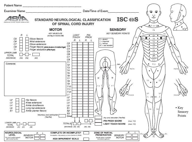
Yoni would love us to document ASIA scores for any spinal trauma patients. This is good practice for physical examination WBAs and fellowship exams.
ECG of the Week
The ambulance call through a signal 1 STEMI call on a 50 yo female with acute and ongoing chest pain. They fax the below ECG.
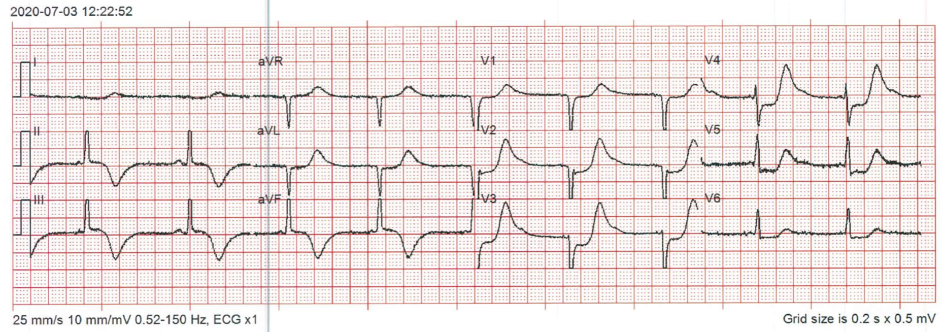
This ECG is concerning for ischaemia although on its own it does not quite meet criteria for a STEMI. There is <1mm STE in inferior leads, deep inverted T waved inferior leads, horizontal ST depression V2-V6. This is concerning for a possible posterior STEMI and would warrant a posterior ECG.
So what are the criteria for a STEMI. Well, this has changed over recent decades and almost certainly will continue to change. The European, American and Australian guidelines are all similar but with some differences.
Currently, the Australian Heart Association Guidelines suggest the following for a STEMI definition:
Clinical history of typical chest discomfort or pain of ≥ 20 minutes duration
And
ECG Criteria with persistent (>20 minutes) ST segment elevation in ≥ 2 contiguous leads of:
≥2.5mm ST elevation in V2/3 in men under 40 years
Or
≥2.0mm ST elevation in V2/V3 in men over 40 years
Or
≥1.5mm ST elevation in V2/V3 in women
Or
≥1.0mm in other leads
Or
Development of new onset LBBB
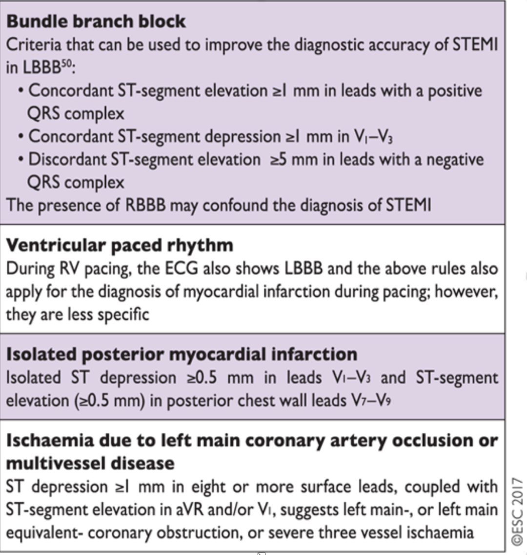
Of note, new LBBB is still in the Australian guidelines although recent evidence has largely refuted this concept and the European and American guidelines are going against this. I suspect this will change in the next guidelines.
The European guidelines, which are also the most recent of the three, also suggest urgent cath lab activation for STEMI equivalents as shown in the following table. I also suspect that this will come in to the American and Australian guidelines soon.
The Australian guidelines are well worth reading and can be found here. I brought up one other teaching point from these guidelines.
When can we do a single troponin in the ED? We are potentially missing important patients when we don’t follow the guidelines. There are only two scenarios when we can do this (using a HS troponin)
Patients whose pain and symptoms resolved 12 hours prior to testing where the cut point is <99th percentile
Patients with symptom onset > 3 hours ago and the troponin is < level of detection of the specific assay (not < 99th percentile)
Images of the Week
It’s 6 am in regional Victoria. A 38 yo female presents after her left foot was trapped under a fork lift.
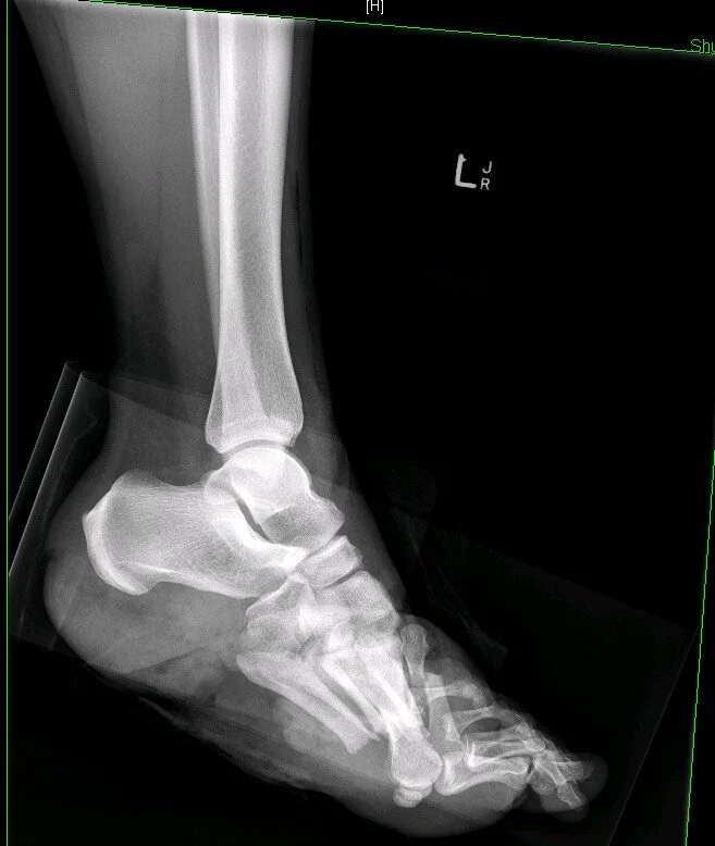
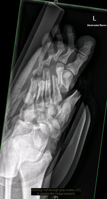
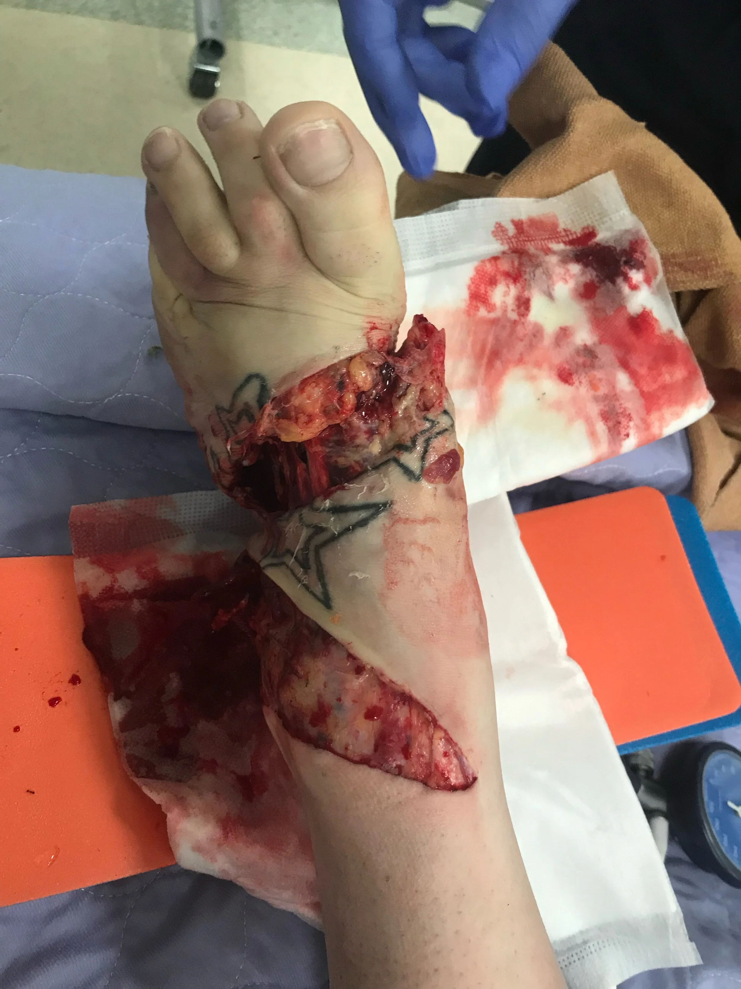
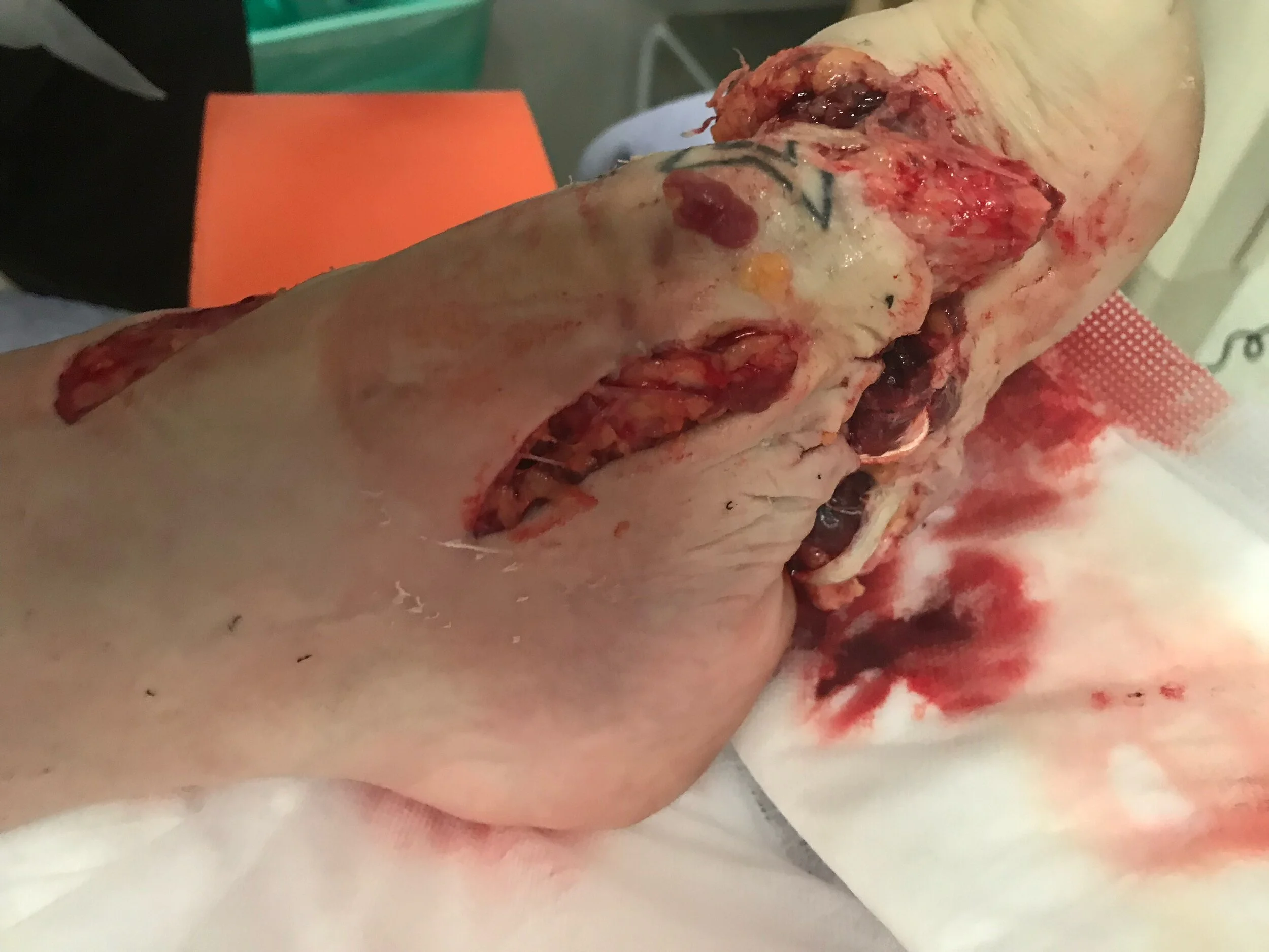
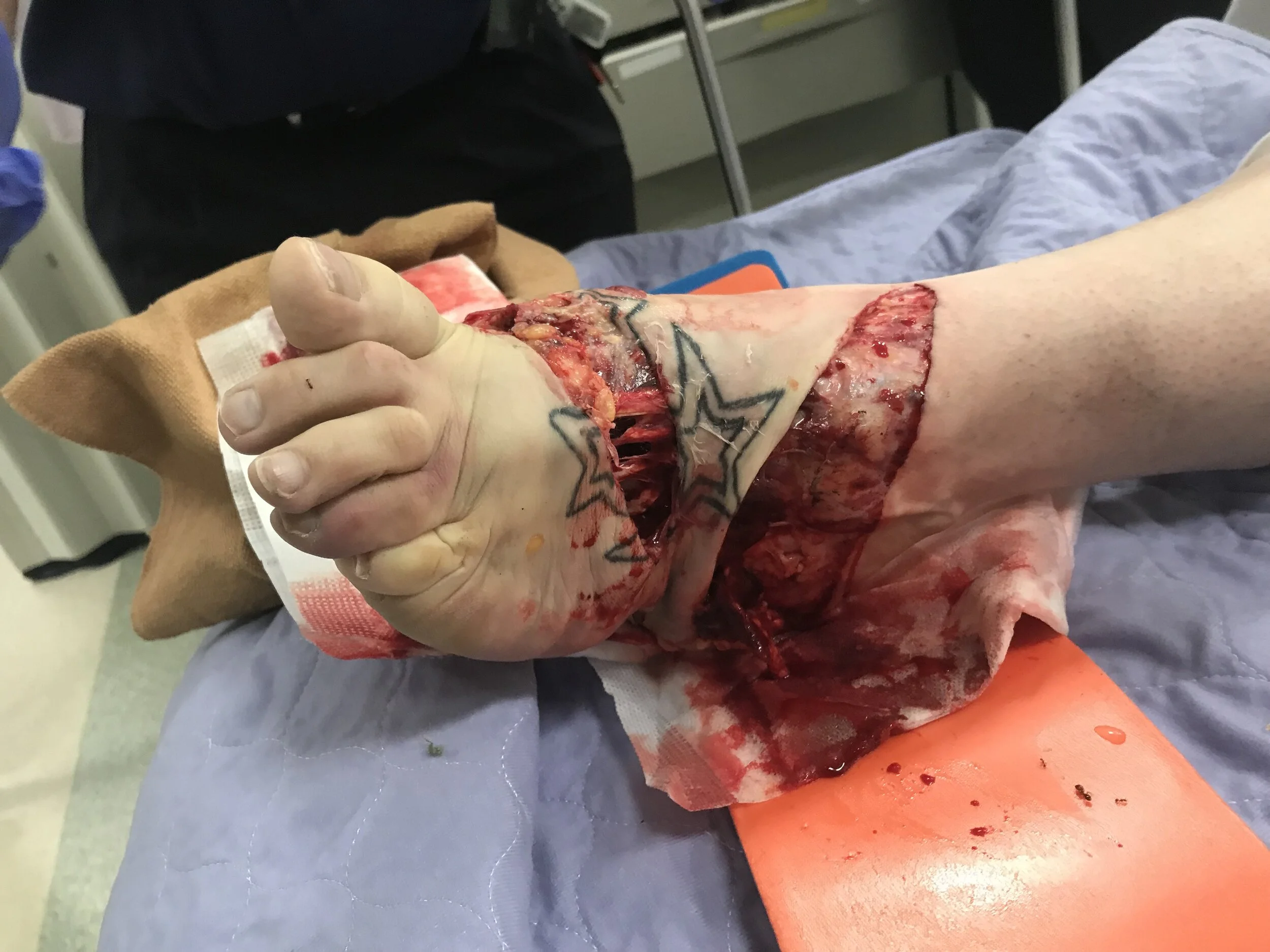
The images show a compound fracture of the mid-foot with a large complex wound. There is active bleeding, tendon and bone visible and likely a protruding 1st metatarsal. The wound is contaminated and there is pallor of the forefoot suggesting vascular compromise.
The xrays show 1st - 5th metatarsal shaft fractures. There are also dislocated 3rd-5th MTP joint dislocations and gas within the soft tissues.
Management should involve haemorrhage control, pain relief, irrigation of the wound (either under sedation in the ED or immediate transfer to theatre), infection prevention with antibiotics and tetanus prophylaxis.
Although attempts were made to salvage her foot she ended up with a mid-foot amputation and then later a below knee amputation.
Gas of the Week
A 76 yo presents with worsening exertional breathlessness over weeks.
SHx: Home alone, indep ADLs.
PHx: OSA on CPAP. Pulmonary HT.
no recent TTE
last sleep study >10 years ago; sats 70-80 when asleep, occasionallhy down to 50’s
No fever, no chest pain
Sats 70% after transfer from chair to bed. Up to 93 on 1l/min NP O2.
Chest clear. Pitting oedema bilat to mid thighs, symmetrical.
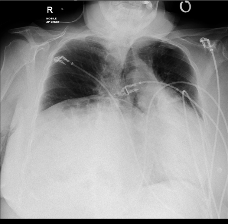
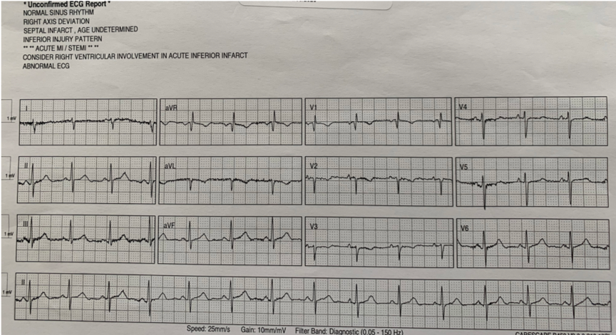
Below are ARTERIAL blood gases on arrival and after a few hours. There is also the patient’s xray and ECG.
Questions
List 5 DDx causes of this person’s respiratory failure
What are the features for/against a PE?
What is the Haldane effect?
Looking at the first arterial gas
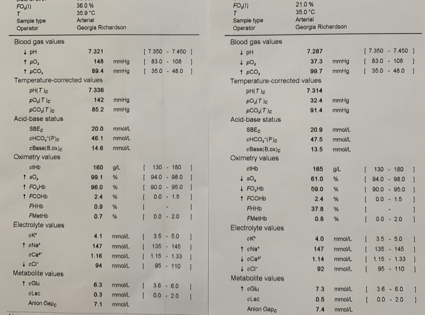
The pH is only mildly acidotic. With a raised pCO2 this suggests a respiratory acidosis.
The expected bicarb in a respiratory acidosis if acute is: 29 if acute or 44 if chronic. With the above given history there is likely a chronic respiratory acidosis with the expected Given the actual bicarb is 46 there is likely also a small metabolic alkalosis present.
If you were to apply Fencl-Stewart approach to this gas then
SID = 147 + 4 - 94 = 57. Therefore the is a hypochloraemic metabolic alkalosis with the effect on the base excess being +15
Lactate effect = 0
Albumin effect unknown but given the patient’s measured base excess is +20 it is possible that this patient’s albumin low.
Now let’s look at the oxygenation
pO2 - patient is hyperoxic on 36% FiO2 (about 4L via NP). Of note the saturations is 99.1%
Aa Gradient = approx 260 - 1.25 x pCO2 (90) - pO2 (148)
= approx 0
Therefore there is no raised Aa gradient and therefore no suggestion of VQ mismatch
So overall, this gas shows us that the patient has a chronic respiratory condition. Even with this, the pCO2 is very high and there is likely an acute component also that overall is not affecting the pH too greatly due to a concurrent metabolic alkalosis. The patient does not have an issue with oxygenation on this gas.
Now look at the gas taken a few hours later after oxygen therapy. The pH is now 7.28, the pO2 37 with sats of 61% and the pCO2 99. The patient has effectively been poisoned with oxygen leading to a decreased respiratory drive and now type 2 respiratory failure.
This is a really good example as to why we should not aim for saturations of 100%, particularly in the patient with possible chronic hypercarbia where the aims should be 88-92%.