
15 July 2020
1000-1030: Recap from last week - Alex, and Gas/ ECG / Image of the week with Alex and Mark
1030-1130: Radiology: Musculoskeletal, commonly missed # : Leon Winata
1130-1145: Break
1145-1200: quick look at SAQ's
1200-1300: New wba forms and assessment : Claudiu
1300-1400: Current situation update with Andrew and Jonty
ECG of the Week
A 34 yo female presents to the ED with progress weakness of her limbs. While in the ED she has repeated episodes of syncope that last several seconds only. One of these happened to be captures on the 12 lead.
In between episodes, HR 80, BP 120/70, Temp 37.0. SpO2 99% RA, RR 16. 1/5 power throughout lower limbs with absent reflexes, 3/5 power in upper limbs
This ECG shows a run of torsades de pointes. Note the broad complex tachycardia with variable amplitude. In the initial part of the rhythm we can see inverted T-Waves followed by prominent U wave.
This patient had a K+ of 1.7
Remember to diagnose Torsades the patient must also have a prolonged QT/QU interval. In the absence of this the diagnosis is likely VT.
There is a good piece on the management of Torsades on EMCRIT -> https://emcrit.org/ibcc/tdp/
In summary,
Treat the underling problem - in this case hypokalaemia
Stop any potential QT prolonging drugs
Give Magnesium - 5-10mmol over 10 minute followed by standard infusions being ware of causing hypotension
In refractory cases, consider speeding up the heart (from EMCRIT)
Speeding up the heart rate will decrease the QT interval and reduce the risk of acquired torsades (but it may be contraindicated in some forms of congenital torsades).
The usefulness of chronotropy depends on the patient's baseline heart rate.
Chronotropy is most beneficial for patients starting out with bradycardia.
If the patient is already significantly tachycardic, chronotropy is unlikely to provide benefit. The usual target heart rate is 100-110 b/m, but occasionally heart rates up to 140 b/m may be needed. There's no high-quality data on this.
Medical chronotropy is generally the easiest & fastest way to stabilize the patient. The ideal chronotrope depends on the patient's hemodynamics and baseline blood pressure:
Baseline severe hypotension: adrenaline infusion.
Baseline normotension or mild hypotension: isoprenaline infusion.
Electrical chronotropy may be used if medical chronotropy fails:
Transcutaneous pacing may work, but this is painful for conscious patients.
Transvenous pacing is more comfortable, but this is more invasive and takes a bit longer to achieve.
Patients with a pacemaker may have the device rate increased.
Image of the Week
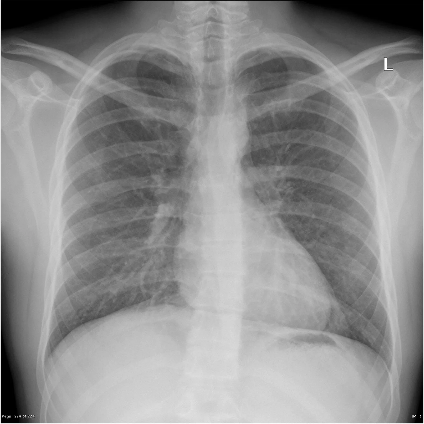
A 23 yo male presents to the ED with sudden onset left sided pleuritic chest pain.
SpO2 99% RA, RR 14
Not unwell looking
Chest clear to auscultation
The following X-ray is obtained.
Case courtesy of Assoc Prof Frank Gaillard, Radiopaedia.org
This xray shows the continuous diaphragm sign. It is subtle but it is not normal to be able to trace the diaphragm all the way across the thorax as you can in this image. This is suggestive of a pneumomediastinum. You can also see a small amount of air between the heart and pericardial fat at the left inferior border of the heart and also around the aortic knuckle.
In most cases the treatment is supportive only. Specific consideration needs to be considered in divers or patients that may travel by air.
Gas of the Week
A 38 yo male presented to the ED with possible alcohol withdrawal. Initially he looked unwell, agitated, tremulous, febrile 38.3, SBP 90 HR 150.
Shortly after the first VBG (at 1615) he had a cardiac arrest (asystole -> PEA -> ROSC after 35 minutes including thrombolysis - given due to a possible dilated right ventricle on ultrasound).
What is your interpretation of the first blood gas at 1615. Now look at the second gas, is a pH of 6.60 with lactate of 28.0 survivable?
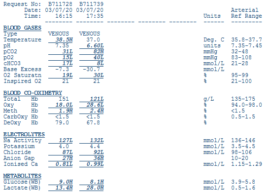
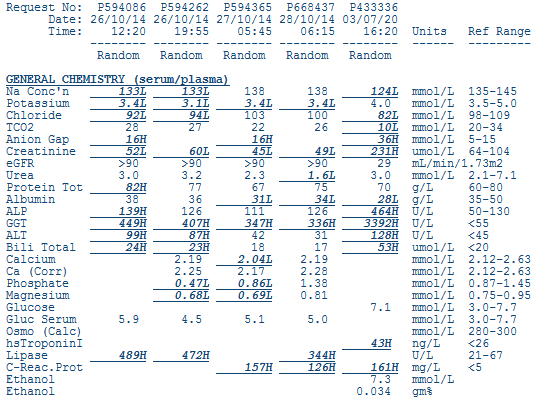
If we look at the first gas the patient has a normal pH. However with a base excess of -7 there must be both an acidosis and alkalosis occurring.
Using Fencl Stewart:
SID = 127 + 4 - 87 = 44. Therefore there is a very mild hypochloraemic metabolic alkalosis. Effect on BE is +2
Albumin effect. (42-28)/4 = 3. Effect on BE is +3
Lactate effect = - 12
Sum effect is -7 which matches the measured base excess
If we use the traditional method:
AG = 127 - 87 - 17 = 23. Therefore HAGMA.
Delta ratio = (23-12)/(24-17) = 1.5 Therefore isolated HAGMA of which lactate is the most likely cause
As you can see the patient’s second blood gas shows a marked deterioration post cardiac arrest. A pH of 6.6 is generally not compatible with survival. This patient unfortunately died in the ICU later that night.