
5 August 2020 - Orientation and COVID
Program
10.00-10.30: Welcome to SVHED, expectations of teaching, DEMT stuff: Sarah
10.30-11.30: COVID: Keeping ourselves safe: PPE/ AGP/ COVERT process / modifications to ALS/BLS: Alex Handrinos
11.30-11.40: Coffee/ freshly baked banana and date muffin
11.40-12.10: Update on COVID: Mark Rugless
12.10-12.40: How to improve your presentations: Alex Handrinos
12.40-12.50: wba’s : Sarah
12.50-1300: Break
13.00-13.30: Journal Club: How to critically appraise a paper: Hamed Akhlaghi
13.30-14.00 ECG/ image/ gas of the week : Alex/Sarah/Mark
Journal Club Article
Jun, J.‐K., Winata, L.S.‐h., Morrissey, B. and Sutherland, T. (2020), Elimination of oral contrast leads to faster scans in the workup of abdominal pain in the emergency setting without compromising diagnostic accuracy. J Med Imaging Radiat Oncol. doi:10.1111/1754-9485.13053
Key Summaries
There was lot’s of orientation type of teaching this week. Please go to Sharepoint on the intranet for all COVID related guidelines, policies, education as this content is changing frequently.
Go to the post on How to Improve Your Presentations to view the recording of this talk and key learning points.
ECG of the Week
An 82 yo NESB patient is brought by ambulance after two syncopal episodes. Unknown medical history.
This ECG shows pacemaker failure. Note that pacing spikes are not always seen in all of the leads. In this case only well seen in the lateral leads although very small spikes can be appreciated in I, aVR and V1 if you look closely.
We can see that the pacemaker is sensing appropriately with pacing spikes following p waves however there is no subsequent ventricular beat. This is therefore failure to capture. Of concern, in the rhythm that we can see there is no underling ventricular rhythm and therefore this patient is pacemaker dependent.
The patient requires urgent pacemaker assessment, treatment of reversible causes and if hypotensive will require transcutaneous or trans venous pacing.
Pacemaker failures can be divided into failure to sense and failure to pace.
Failure to sense
The pacemaker fails to sense an intrinsic rhythm potentially causing a dangerous R on T phenomenon
This is rare with modern pacemakers
Failure to pace
This could be failure to output. No pacing spikes are seen and can be due to battery failure (rare), lead problems or the pacemaker oversensing such as with muscle fasiculations
It could also be due to failure to capture as in the case above. Here, pacing spikes are seen but the myocardium is not ‘captured’ and no ventricular beat occurs. Causes include MI at the site of the lead (often RV), endocardial fibrosis, drugs such as amiodarone, hyperkalaemia and pacemaker failure from battery/lead problems.
Image of the Week
A 52 year old male presents with an inversion injury to left ankle earlier today. He had mobilised on it initially but it then painful several hours later. On examination there was slight bruising over the lateral collateral ligament. PMH: AMI and PTCA 6/52 ago.
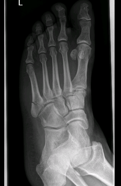
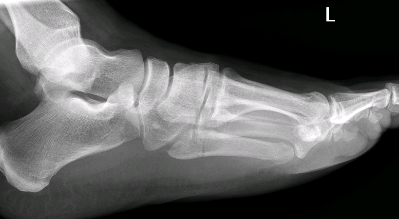
These X-rays demonstrate a Jones Fracture.
Typically these fractures are located at the metadiaphyseal junction approximately 2cm from the tip of the 5th metatarsal. They are predominantly horizontal and should not extend distally or to the articular surfaces.
This is an important one to recognise as the complication rate of non-union are high. Internal fixation or bone grafting is sometimes necessary and healing can take greater than two months due to the watershed vascular supply to the area.
These patients require non weight bearing immobilisation and referral to an orthopaedic surgeon.
Differential diagnoses
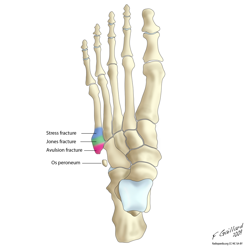
Avulsion fracture: Sometimes known as a pseudojones fracture. Proximal, treated conservatively.
Os peroneum: well corticated bone. Seen in 25%
Stress fracture: often more distal - usually about 1.5cm from the metadiaphyseal junction.
Normal apophysis of the fifth metatarsal: Lies laterally and oriented longitudinally.
Gas of the Week
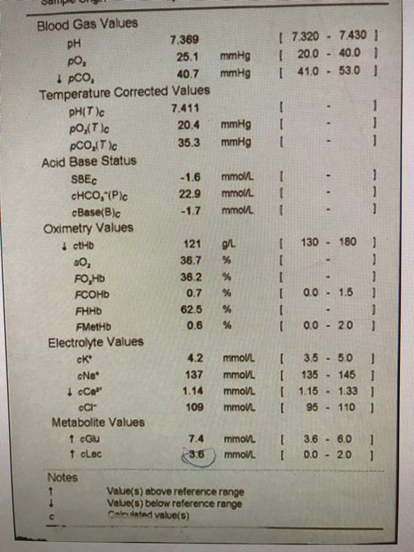
43 yo female with anorexia nervosa and alcohol misuse. She was last seen to eat two weeks ago. For the past four days she has been bed-bound and faecally incontinent in bed. When her husband tried to get her up to have a shower she lost consciousness whenever in the upright position. Initial BP 60 systolic unproved to 100 systolic with auto infusion. She was confused and had pitting oedema to the knees.
The HMO brings you the following gas surprised at how normal it is.
Is there a major problem here?
You assess her to be intravascularly volume deplete. What fluid do you choose to give and why?
This gas is unlike to come up in the exam.
pH normal, bicarb normal, base excess normal and all electrolytes are in the normal range. So why isn’t this just a normal gas?
Fencl-Stewart approach is clearly the better method here.
SID = 137 + 4 - 109 =32. Therefore there is a hyperchloraemic acidosis with the chloride effect on base excess -10
Lactate effect on base excess is -2.6
Albumin effect unknown as albumin not given
The sum effect on base excess is -12.6. The measured base excess is rounded to -2 so there must be another alkalotic process contributing +10 to the base excess
There is not much that contributes to an alkalosis that is not measured in the strong ion difference. But what about the albumin? Remember that albumin must drop by 4 for every 1 increase in base excess. So in this case it would need to drop by 40!!
Well, the patient’s measured albumin was 6. This patient is therefore extremely unwell.
In regards to interpretation, such a low albumin is likely nutritional in this case +/- a catabolic process. When you look at the causes of a hyperchloraemic metabolic acidosis (commonly described as a NAGMA) it is probably due to diarrhoea or renal tubular acidosis.