29 July 2020 - Ophthalmology
Program
10-1015hr – Recap and welcome
1015-1045hr – The Red Eye – Luke
1045-1115hr – Lateral canthotomy – Alex H
1115-1130hr – coffee break
1130-1200hr – ECG/gas/image of the week
1200-1215hr – Ten Min Tox – Jacqueline
1215-1245hr – Eye SAQs – Sarah
1245-1300hr – quick lunch break
1300-1400hr – Disaster! Andrew Walby
Key Learning Points
The Red Eye
There are plenty of causes of the red eye and an approach including a good history and examination is vital. Here are some pearls when thinking about the red eye.
Gonococcal conjunctivitis causes copious discharge. You wipe it away and by the time you are done there is more! Yuck.
A dendritic ulcer suggests a herpetic infection but be aware that a herpes infection can be present without any ulceration
UV keratitis presents hours after the actual burn and typically has punctate lesions on slit lamp exam
Consensual photophobia is a key finding in iritis. That is, pain in the symptomatic eye when the unaffected eye is exposed to light
Always consider herpes zoster ophthalmicus in any patient with a facial rash or facial pain
Contact lens wearers can get keratitis. Remove and throw out the contact lenses.
Lateral Canthotomy and Cantholysis
This is a low frequency, high acuity procedure that we all need to be able to do. It is relatively straightforward, has minimal morbidity associated with it and can be sight-saving.
There are two procedures. A lateral canthotomy (incision of the lateral cants) which is followed by a cantholysis (cutting of the inferior canthal tendon) that are performed in sequence in patients with suspected acute orbital syndrome. Usually, these are patients who have had trauma to the face and have a retrobulbar haematoma. This is relatively rare however this haematoma leads to damage to the retinal artery and optic nerve with the critical ischaemic time being about 60-90 minutes.
We reviewed a video of the procedure being performed. There are plenty of links online.
Indications
Unconscious patients: relative afferent pupillary defect (RAPD) is the most reliable sign or an intraocular pressure (IOP) of >40mmHg
Conscious patients: Visual acuity < 6/18 and one of IOP >40mmHg or RAPD or proptosis
When in doubt, almost always better to perform the procedure.
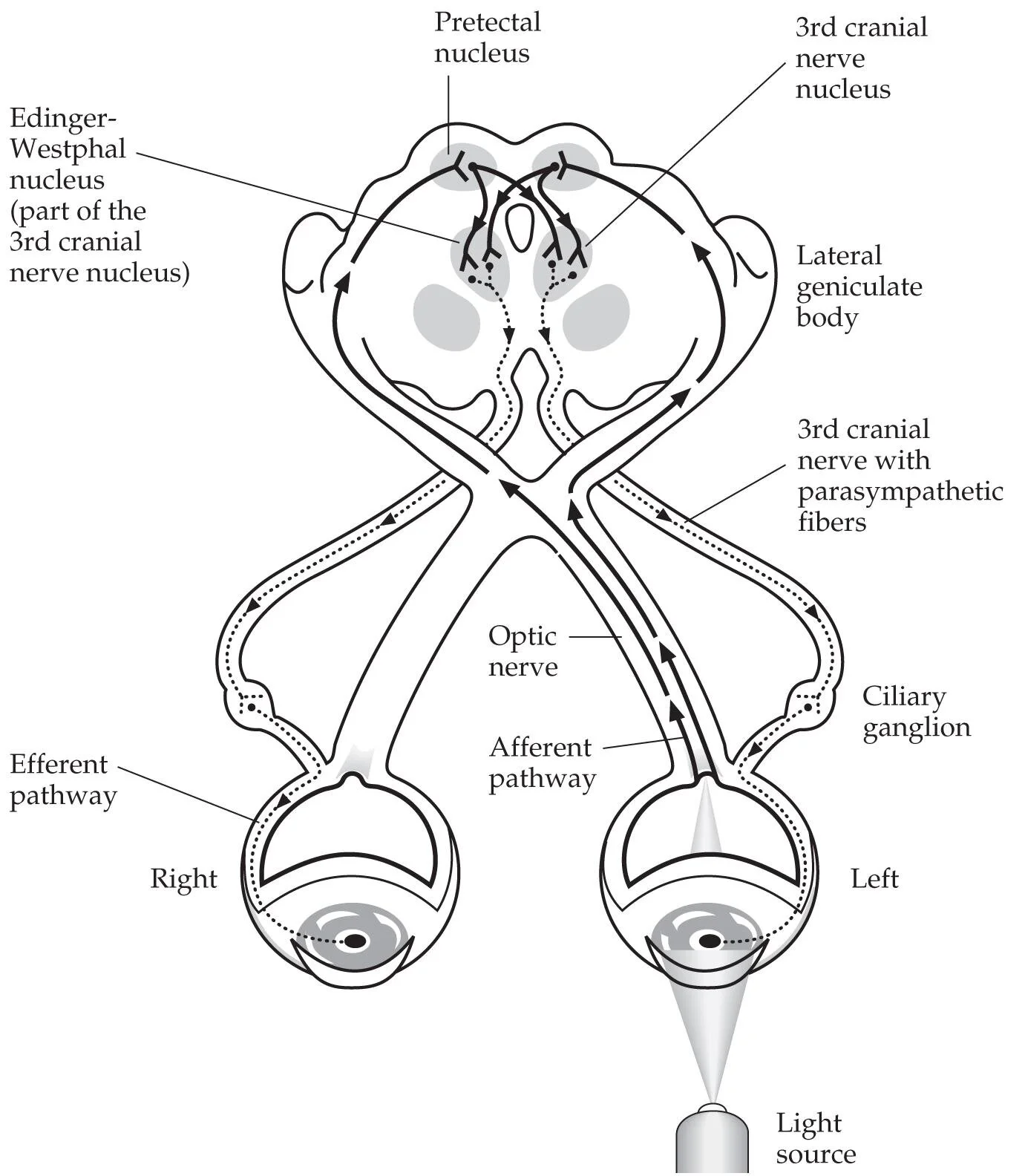
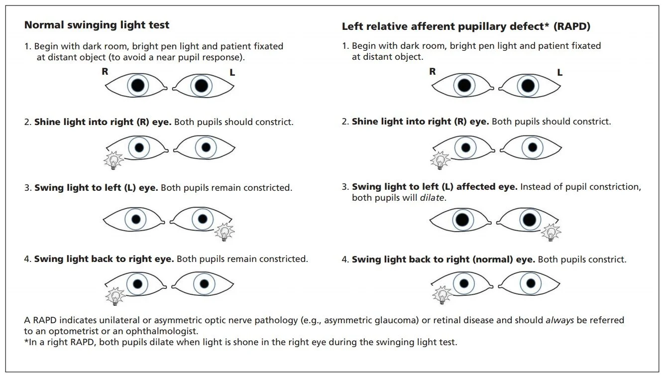
RAPD
We reviewed how to examine for an RAPD which is often a sign of optic nerve dysfunction. Also commonly caused by optic neuritis, ischaemic retinal disease, retinal detachment, optic nerve damage from trauma/radiation/tumour.
Toxicology - Organophosphate Poisoning
Typically causes a cholinergic syndrome
Nicotinic: Tachycardia, hypertension, fasciculations, muscle weakness
Muscarinic: salivation, bronchorrhoea, lacrimation, diarrhoea, miosis, bradycardia, hypotension, vomiting
These patients die from bronchorrhoea
The antidote is atropine
Give 1200mcg IV then double the dose every five minutes until:
Adequate atropinisation: HR >80, BP > 80, chest clear
Pralidoxime may be beneficial: discuss with a toxicologist
ECG of the Week
An 80 yo ex-smoker presents with cough, dyspnoea and wheeze.
The rhythm is irregular, narrow and fast and is therefore one of: AF, atrial flutter with variable block or multifocal atrial tachycardia. There are P waves preceding every QRS however they have variable morphology suggesting they originate from different parts of the atria. Therefore this ECG shows multifocal atrial tachycardia (MAT).
The ECG also shows a right axis, dominant R wave in V1 deep S wave in V6 which are consistent with cor pulmonale.
MAT is most commonly seen in advanced CCF and COAD. There is no specific treatment other than to treat the underlying cause. The arrhythmia itself does not usually cause significant physiological disturbance.
Image of the week
A 75 yo NESB Vietnamese male is seen in the COVID zone of the ED. He was admitted under the COVID unit last week, is known COVID positive, and then discharged home but has not managed well and has represented.
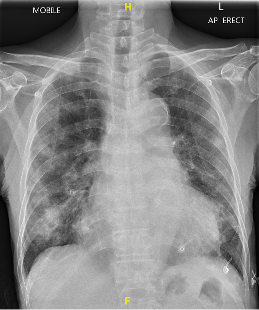
This CXR shows patchy consolidation in both lower zones. There is an irregularity in the right lower zone that is suspicious for a cavitating lesion. In this NESB patient from Vietnam it brought up a concern for active tuberculosis. Imagine that, TB and COVID at the same time!
Thankfully, it turned out to not be TB and with that went my case write up.
A reminder as per our hospital policy on imaging in COVID. CXR should not be used to diagnose COVID. Only 70% of patients requiring hospital admission have X-ray findings at the time of admission. It is suggested that we X-ray patients who we are suspicious might have a lower respiratory tract infection (think hypoxia, particularly unwell, or crackles if you haven’t ditched your stethoscope yet). If the patient just has upper respiratory tract symptoms then X-ray is probably not that useful. I liken this to the child with bronchiolitis. We only really x-ray them if we are suspicious of pneumonia.
Typical x-ray findings include bilateral, peripheral and lower zone airspace opacities similar to atypical pneumonia.
Gas of the week
The same patient as above had the following vital signs: HR 95, BP 140/80, temp 36.5, RR 28. He was cachectic, volume deplete and had cold peripheries. Unable to obtain a SpO2 trace using any method - he did not appear cyanotic but had relatively dark skin so difficult to assess. An arterial gas was taken to assess oxygenation on room air at the time. (Albumin 34)
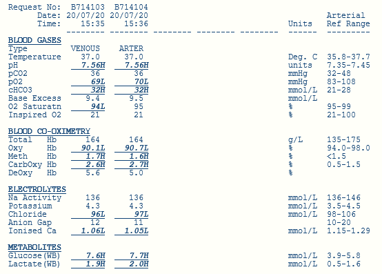
This is an interesting gas.
There is a primary metabolic alkalosis as determined by a high pH, high HCO3 and high base excess.
Is there a secondary respiratory acid base disturbance? Expected PCO2 = 0.7 x HCO3 + 20 = 42. With the actual pCO2 36 there is a mild respiratory alkalosis present. Respiratory alkalosis is almost always caused by hyperventilation from whatever cause.
Looking at the metabolic state in further detail:
Fencl-Stewart:
SID = 136 + 4 - 97 = 43, therefore minimal chloride effect on the base excess of +1
Albumin effect = (42-34)/4, therefore albumin effect on the base excess is +2
Lactate effect = -1
So the sum effect is + 2 yet the actual measured base excess is +9.5 so there must be another alkalosis occurring. Mark discussed whether this could be post hypoventilation syndrome?
Traditional approach
Traditional exam teaching would be that AG and delta ratio are not required given the process is an alkalosis.
AG = 7 (or 11 if using potassium)
Causes of a metabolic alkalosis using the acronym GROE
G - GI Acid Loss - Vomiting (most common), diarrhoea, Ileostomy
R - Renal Acid loss - Diuretics, Bartter/Gitelman syndrome, milk-alkali syndrome
O - Overdose of base - antacids, laxatives, iatrogenic HCO3 administration
E - Endocrinopathy - Steroid excess, Cushing syndrome