
12 August 2020 - Cardiology
1000-1030: Welcome, interesting case presentation: Sarah, Wellness update Mim
1030-1130: Exercise Induced Arrhythmias: Neil Cunningham
1130-1140: Break, stretch, another coffee
1140-1210: Low risk Chest Pain: Mim - Click here for handout with reference tables
1210-1240: Bradyarrythmias: Khalid
1240-1250: Break
1250-1320: ECG/ Image/ Gas of the week: Sarah, Mark, Maria
1320- 1400: SAQs: Sarah
Key Points
Exercise Induced Arrhythmias
The most common causes of sudden cardiac death in athletes
HCM
Anomalous coronary artery
Acute aortic syndrome - dissection or rupture
Exercise related syncope, chest pain or dyspnoea either during the exercise or up to 30 minutes after are red flags and should prompt further cardiac investigations. These patients must be advised to avoid heavy exertion until further workup is able to be completed.
Normal ECG findings in athletes include:
sinus bradycardia
sinus arrhythmia
ectopic atrial/junctional escape rhythm
1st degree or Wenckebach AV block
incomplete RBBB
early depolarisation
The Adult Pre-Screening Tool that Neil discussed can be found here.
Bradyarrhythmias
1st degree HB and Mobitz Type 1 HB are relatively benign
Common causes of heart block include:
Ischaemia: especially inferior MI
Degenerative heart disease
Drugs: BB, CCB, Digoxin
Hyperkalaemia
Treatment of bradycardias in general can include:
Removal of the contributing agent
Optimise electrolytes
Atropine 600mcg IV - often ineffective unless increased vagal tone
Isoprenaline infusion (I often start at 3mcg/min and titrate every 5 minutes). Can also use adrenaline.
Transcutaneous pacing
Low Risk Chest Pain
What we, as clinicians, consider low risk chest pain may differ from what a patient considers low risk.
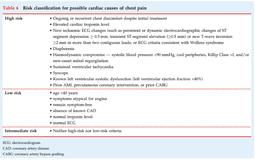
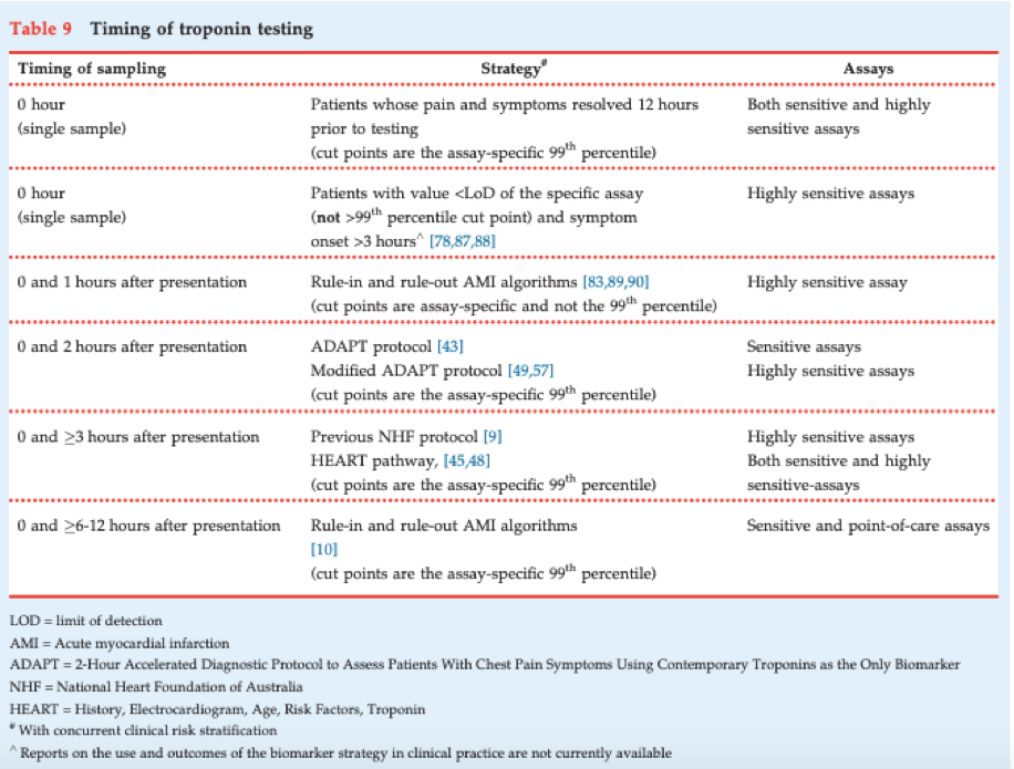
ECG of the Week
A 48 yo man walks into a tertiary ED with ongoing crushing central chest pain for the past 60 minutes. He has cardiac risk factors: smoking, family history, hypertension. His BP is 130/80. GCS 15. RR 28. SpO2 99% RA. Here is his ECG.
This ECG is a good example of a high lateral STEMI. Note the ST elevation in high lateral leads I and aVL and reciprocal ST depression in the inferior leads.
For some reason, we easily miss these STEMIs by not considering leads I and aVL. When we see ST depression in inferior leads, rather than assume this is an inferior NSTEMI or ischaemia, we need to actively seek ST elevation in the high lateral leads.
Image of the Week
A 36 yo female is brought by ambulance with a blood pressure of 60/40 which is not fluid responsive. Her other observations are within normal limits. She is complaining of right sided chest pain and RUQ pain. She reports her only medical history to be a cholecystectomy 5 years ago.
A slice of her CT is shown below.
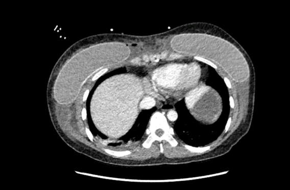
This slice of the CT shows axillary soft tissue inflammation and gas locules around a silicon breast implant, raising the suspicion of a necrotising soft tissue infection.
Learning points from discussion:
Think necrotising soft tissue infection where there is disproportionate pain or rapid clinical deterioration
Necrotising fasciitis is a surgical diagnosis; if there is any suspicion these patients need urgent referral to plastics and a surgical incision to confirm the diagnosis and debride infected tissue
Most are polymicrobial in at-risk patients; infections in young healthy patients tend to be related to virulent organisms such as group A strep, MRSA and clostridium perfringems
Empirical treatment is meropenem or taz + vanc; many ID specialists also recommend adding clindamycin as it has a static effect on exfoliative toxin production
Beware of confirmation bias and always re-examine the patient if the imaging doesn’t support your initial diagnosis
Look at the whole image systematically and think outside the ‘box’ for CTs – don’t forget the soft tissues
Gas of the Week
A 70 yo male was transferred from a regional hospital for specialist vascular surgery care. He had recently been admitted to St Vincent’s with a DVT and PE before being transferred back to the regional hospital. He developed a psoas haematoma with active bleeding in the setting of anticoagulation (clexane) and was transferred back to St Vincent’s. To make matters worse he was also being treated for candida sepsis. He underwent interventional radiology. While in hospital he picked up COVID and then died with respiratory failure. Wow.
Here is his arterial gas (9/7/20 at 2120) taken shortly after arriving to St Vincent’s.
How many acid-base disturbances are going on here?
What are they?
What formula do you use to determine expected PCO2 in this patient?
The primary disturbance is a metabolic acidosis based on the negative base excess, low bicarb and low pCO2. Overall there is only a subtle acidoses present due to respiratory compensation.
Traditional method
AG = 134 - 103 - 11 = 20 (or 25 if including the potassium). Therefore there is a HAGMA
Delta ratio = (20-12)/(24-11) = 8/13. This indicates an additional NAGMA as well.
Fencl-Stewart method
SID = 134 + 5 - 103 = 36. There is therefore a hyperchloraemic metabolic acidosis with the effect on the base excess of -6
Lactate effect = -9
Albumin effect = + 4
Sum effect = -6 + (-9) +4 = -11
The measured base excess is -13 so perhaps there is a small effect of ketones but overall this is within the margin of error of +/- 2
Expected PCO2 = 1.5 x 11 +8 = 24. The actual pCO2 is 20 so there is also a very small respiratory alkalosis - this might be within the margin or error or there might also be pain/anxiety/delirium etc.
A-a gradient = 150 - 1.25 x 20 - 100 = 25. There is no significant VQ mismatch given that normal A-a gradient increases with age. (1-2mmHg for every decade). So although a PE is mentioned in the stem, it is not particularly significant here.\
Clinical interpretation
There are multiple processes here
Hyperchloraemic metabolic acidosis - likely RTA
Lactic acidosis - ? tissue hypoperfusion, ? abdominal compartment syndrome
Hypoalbuminaemic alkalosis - catabolic process, nutritional deficiency
Respiratory alkalosis - hyperventilation from pain, anxiety, delirium