
19 August 2020 - Respiratory
Program
1000-1015hr – Welcome, Recap - Maria
1015-1100hr – When COVID arrived in the ED - DOM presentation, Jonty
1100-1130hr – Update on COPD/Asthma – Maria
1130-1145hr – coffee break
1145-1215hr - ‘Lung infections’ – Phil
1215-1245hr – ECG/image/gas – Alex and Maria
1245-1300hr – lunch break
1300-1315hr – Joel – Registrar forum
1315-1400hr – SAQs – Jacqueline
Key Learning Points
Asthma/COPD
Treatment of life-threatening asthma includes:
Bronchodilators - continuous nebulised salbutamol, add ipratropium
Remember in COVID times can give 12 puffs of salbutamol and 8 puffs of ipratropium via pMDI and spacer, repeated doses
IV salbutamol can be used but has a high side effect profile and many are unfamiliar with its use.
Steroids - dexamethasone 8mg IV or hydrocortisone 200mg IV. Give these early but note they won’t work for hours
Oxygen - titrated to SpO2 >94%
Adjunctive medical therapies
Magnesium - 10mmol IV over 20 minutes
Aminophylline - 500mg IV
Dissociative ketamine - but be prepared to intubate
Adrenaline - 500mcg IM or low dose IV push doses (i.e. 10-20mcg). Remember that anaphylaxis can look very much like life threatening asthma.
Non-invasive ventilation - controversial and mixed evidence to date
Intubation - critical event with high risk of peri-intubation arrest.
Once ventilated need to use careful ventilation (low PEEP, I:E ratio >1:4, low RR, permissive hypercarbia)
COPD - remember to aim for SpO2 88-92% only. it can be very easy to leave the patient for an hour with too much oxygen and return to find them near respiratory arrest.
Respiratory Infections
Strep pneumonia is always top of the list of organisms most likely to cause pneumonia. Other pathogens most likely to cause death in CAP include staph aureus, legionella and pseudomonas aeruginosa
If working in far north Australia, or returned travellers we need to consider Burkholderia. These patients need meropenem when unwell.
POCUS signs consistent with pneumonia can include:
Early: B-lines and and small areas of sub pleural consolidation
Late: Hepatisation of lung, Shred sign
LITFL has nice examples of these https://litfl.com/lung-ultrasound-pneumonia/
ECG of the Week
A 74 yo female presents with acute dyspnoea and diaphoresis that began 2 hours ago. She is triaged to the COVID zone and the following ECG is taken.
Vitals: HR 70, BP 110/90, RR 22, SpO2 95% RA, Temp 36.0
PMHx: PPM, hypertension, ex-smoker.
Meds: Perindopril, atorvastatin
This ECG shows a ventricular paced rhythm. There is one native beat (#5). There are also p waves seen without QRS complexes in the rhythm strip suggesting an underlying heart block.
There is ST depression in V2-V6. With this history as given above this is concerning for an acute coronary syndrome requiring urgent reperfusion. Of note, the ST elevation in the inferior leads is not considered inferior ischaemia as this is normal with a ventricular paced rhythm and a negative QRS complex.
Interpreting a paced ECG for ischaemia is challenging. It has been suggested that Scarbossa Criteria can be used here. The European Society of Cardiology guidelines include Scarbossa for paced rhythms but the American and Australian do not (yet ?) include them.
Modified Scarbossa criteria
≥ 1 lead with ≥1 mm of concordant ST elevation
≥ 1 lead of V1-V3 with ≥ 1 mm of concordant ST depression
≥ 1 lead anywhere with ≥ 1 mm STE and proportionally excessive discordant STE, as defined by ≥ 25% of the depth of the preceding S-wave.
Image of the Week
77 yo male, BIBA with an altered conscious state.
His neighbour called police for a welfare check as he had failed to bring in his bins. Found seated on his couch, incontinent of urine. GCS 12. Examination revealed a left sided facial droop and weakness in the left arm and leg. Unknown how long he had been unwell.
On arrival to ED began having runs of VT and also intermittent partial seizures.
Hypertensive, 180/90. HR 100. SpO2 95% RA. RR 12. BSL 25. Temp 35.0
Code stroke called, taken to CT with the below image obtained. Patient usually takes apixaban for AF.
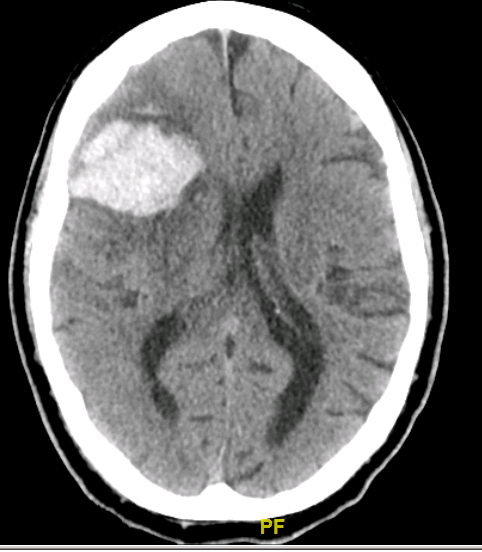
This CT shows a large right frontal intraparenchymal haematoma. There is associated sulcal effacement, midline shift of 5-10mm and sub-falcine herniation.
The most common causes of a bleed like this is hypertension and amyloid angiopathy.
Gas of the Week
An 88 yo male BIBA with falls. Police attended for a welfare check after his neighbour noticed he didn’t bring in the bins. They found him on the ground with a head injury. Subsequent CTs showed facial bone fractures but nil else.
PMHx: HFrEF, Prostate Ca, OA, Cognitive impairment
Meds: aspirin, bicalutamide, ISMN, nebivolol, perindopril, spironolactone, paracetamol
The patient presented 12 hours prior to these bloods tests (this is the first blood gas). His initial biochemistry showed Na 146, K 5.8, Chloride 117. He was given 10 units of actrapid and 50mls of dextrose followed by 1 litre of 5% dextrose overnight. No other IV fluids or treatment given. Here are the morning bloods and gas.
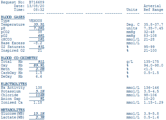
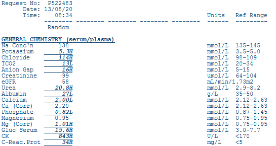
Primary disturbance
- Metabolic acidosis, low base excess, low bicarb
Secondary disturbances
Respiratory compensation – cannot assess as venous gas
Traditional Method
AG = 138 – 116 – 18 = 4 (9 if inc K)
Therefore NAGMA
Fencl-Stewart
SID = 138 + 5 – 116 = 27. Therefore there is a hyperchloraemic metabolic acidosis. Effect on BE is -15
Lactate effect = -1
Albumin effect = +4
Sum effect = -12
Measured effect is -8 so there is another mild metabolic acidosis – likely ketones.
Oxygenation
Cannot assess venous sample
Electrolytes and others
Hyperglycaemic
High urea with normal creatinine
Hyperkalaemia ? (mild) – expected potassium for pH = 5.0 +0.5 = 5.5
Raised CK
Clinical interpretation
Likely intravascularly volume deplete – needs fluids despite known heart failure.
NAGMA/Hyperchloraemic acidosis – causes:
A) Adrenal Crisis/Addisons
B) Bicarbonate Loss – diarrhea, renal tubular acidosis, fistulas
C) Chloride – Excessive NaCl
D) Drugs – acetazolamide, spironolactone