
26 August 2020 - Psychiatry
10-1015hr – Welcome and recap – Andy K
1015-1115hr – Morbidity and Mortality – facilitated by Kirsten Cassidy
1115-1130hr – coffee break
1130-1200hr - Jacqueline Morel: the suicidal patient
1200-1230hr – Joel Wilson – Hallucinations, delusions, acute psychosis
1230-1300hr – Gas, ECG, image of the week – Maria, Alex and Mark
1300-1315hr – lunch break
1315-1400hr – SAQs –vs primary viva practice Sarah/Jac
Key Learning Points
Suicidal Patient
Many suicidal individuals see a physician shortly before their death.
An ED visit for suicidal thoughts or behaviours represents a crisis and an opportunity to provide assistance.
Intoxication should not preclude taking a history, but the provider should repeat the interview when the patient is sober.
There is no evidence to suggest that directly asking about the presence of suicidal ideation or intent creates the risk of suicide in people who have not had suicidal thoughts or worsens the risk in those who have.
Psychosis
Defined as the distortion of loss of contact with reality, without clouding of consciousness
Schizophrenia is usually first diagnosed in the teenage years.
First onset of psychosis in middle age should be considered organic until proven otherwise
In the behaviourally disturbed patient, staff safety should always come first.
Don’t physically restrain a patient without chemical restraint.
ECG of the Week
A 68 yo male presents with intermittent chest discomfort. Vital signs are normal.
PMHx: IHD with previous AMI, T2DM, Smoker
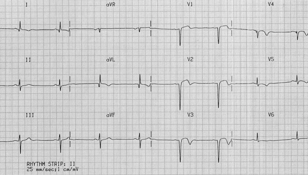
This ECG is an example of an LV aneurysm.
There is minor ST elevation in V1-V3 without any reciprocal ST depression. There are also deep Q waves in V1-V3 with inverted T waves consistent with this patient’s history of a previous myocardial infarction.
LV aneurysm are one of the differential diagnoses for ST elevation in the anterior leads that we often don’t consider. It can be difficult to distinguish from an anterior STEMI but typically there are no reciprocal changes, the ST changes are not dynamic (i.e. they don’t change on repeat ECGs), and there are Q waves indicating prior MI and myocardial scarring. The ST elevation can be concave or convex.
Why care about this diagnosis? Patients with LV aneurysms are at risk of sudden cardiac death, CCF and mural thrombosis and embolism,
Image of the Week
A 28 year old, right-handed lawyer self-presents to ED after getting his tyres stuck in tram tracks and falling off his bike. He complains of right shoulder pain.
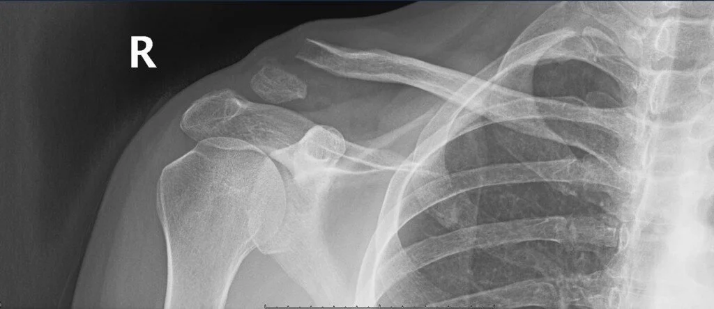
This xray shows a completely displaced distal clavicle fracture
Key points:
Coracoclavicular (CC) ligaments are the key stabilisers of the clavicle
Key neurovascular relations are the divisions of the brachial plexus which run under the middle third, and the subclavian vessels which are deep to the proximal third
Middle third fractures are commonest and generally don’t need fixation, but discuss with ortho if there is > 100% displacement and > 2cm shortening
Distal clavicle fractures need to be referred to orthopedics if there is any loss of integrity of the CC ligaments, which will appear as significant superior displacement of the proximal fragment
Long-term complications include non-union and AC joint arthritis
Gas of the week
An 82 yo greek lady presented with altered bowel habit. She normally has chronic diarrhoea but now presents with BNO for 5 days.
She is complaining of intermittent abdominal discomfort, relieved by flatus. Ie. she is not obstipated*.
PMHx: caecal adenocarcinoma – previous hemicolectomy, T2DM, Chronic kidney disease, frequent infections (UTI/pneumonia/joint infections)
* Obstipation - a complete, severe form of constipation where the patient cannot pass stool and cannot pass flatus.
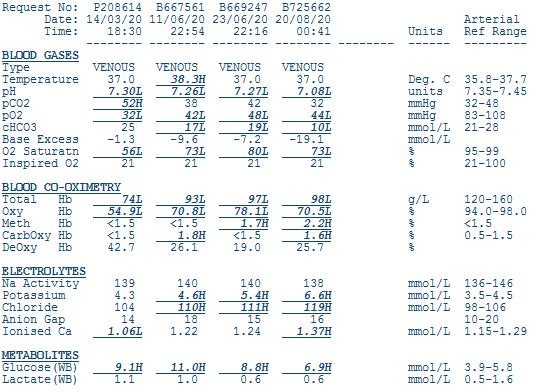
Albumin = 34
Interpretation
There is a moderate to severe metabolic acidosis (low base excess, low bicarb, low pH)
Using Fencl-Stewart
SID = 25, there is a hyperchloraemic metabolic acidosis contributing -17 to the base excess
Lactate effect = 0
Albumin effect = Contributes + 2 to the base excess
The sum effect is -15, the measured base excess is -19 so there must also be another metabolic acidosis from unknown weak acids such as ketones
Using a bicarb-based approach
AG = 15 if including the potassium or 9 if not including the potassium - either way, suggests a NAGMA
Can’t comment on oxygenation or respiratory compensation as this is a venous sample
For every 0.1pH below 7.4 the potassium should rise by 0.5mmol above 5. Therefore the expected potassium with this pH is 6.5. Another way of looking at this is the ‘corrected’ K is 5.1. The patient still has a relative total body potassium excess (probably from CKD) but the measured hyperkalaemia is likely due to the acidosis. Mark suggests treating this patient with sodium bicarbonate.
Clinical interpretation
Like last week, we need to look at the causes of a hyperchloraemic metabolic acidosis, or NAGMA if you prefer the bicarbonate method. With this history, the likely diagnosis is renal tubular acidosis which is almost universal in patients with chronic renal impairment.