
3 June 2020 - Mixed Session
Program
***Geriatric program postponed***
10am-11am – Richard – Delirium
11am-12pm – Ken – Acute assessment of the older person and frailty
12pm- 1pm – lunch and ED stuff (recap from last week, ECG, ABG and image of the week)
1pm-2pm – Min – Geriatric Medicine and community services at St Vincent’s
1pm-3pm - OSCE Group - Brendan
Key Learning Points
With the geriatric program occurring in future weeks we had a few last minute case presentations.
Paediatric status epilepticus
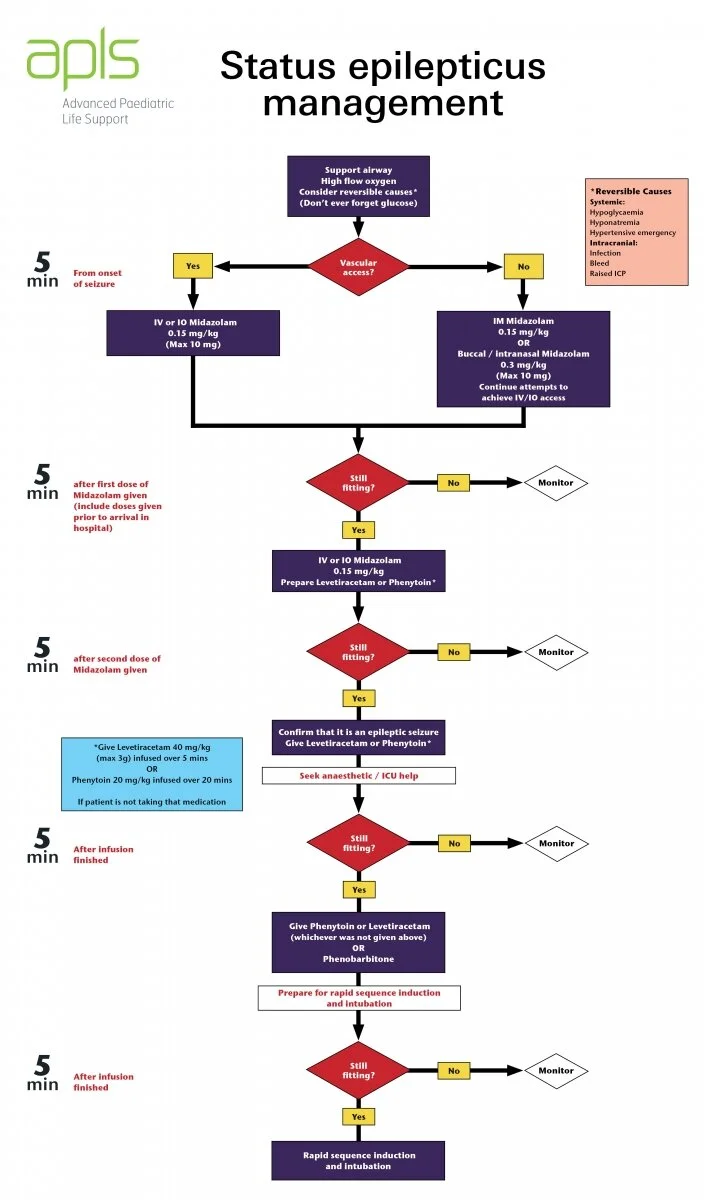
We discussed a case of a two year old who was rushed into The Alfred ED (another adult only department) with status epilepticus. It is worth considering how we would manage this here at St Vincents. Key learning points were:
Support the father, allocate a team member to be with dad
Know the algorithm (or download the APLS app)
Benzo, benzo, antiepileptic, antiepileptic, RSI
See the algorithm attached
Call PIPER early to facilitate management and transfer
Pulmonary Oedema
We also discussed a case of pulmonary oedema post near drowning.
It is worth considering that there are multiple differential diagnoses for pulmonary oedema.
Not only is there cariogenic pulmonary oedema (and its many causes) but also other categories including:
Neurogenic (ICH, seizures, ECT)
Increased capillary permeability (ARDS, near drowning due to surfactant loss)
Drug-induced (opiate overdose, salicylate overdoes)
Raised pulmonary arterial pressure (high altitude, massive PE, air embolism)
Negative pressure pulmonary oedema (airway obstruction, re-expansion post drainage of large effusion)
ECG of the Week
A two year old is brought to the ED after a syncopal episode in the setting of mild gastroenteritis. As part of the workup, the nurse brings you this ECG.
This ECG is normal for a two year old. Findings include a heart rate of about 115bpm, dominant R waves in V1-V3, RSR’ pattern (partial RBBB) in V1 and juvenile T-wave inversion in V1-V3.
The key learning point from this week was to recognise the normal differences between an adult and paediatric ECG. This is all based on physiology where there is right ventricular dominance which is gradually replaced with left ventricular dominance as the child ages. This gives a picture reminiscent of right ventricular hypertrophy in the adult.
Normal findings compared to adults include:
- Right axis deviation (resolves by about 6 months of age)
- Dominant R wave in V1
- T-wave inversions in V1-V3 (Juvenile T waves)
- Shorter conduction periods (PR interval, QRS duration) due to smaller cardiac size
- Sinus arrhythmia
- Higher heart rate
A bit on the precordial T-waves in children:
- For the first week they are upright
- After week one they are inverted (juvenile T-waves)
- They usually become positive again by age 6-8 although can persist into adolescent and early adulthood and this is known as persistent juvenile T-waves
Image of the Week
A 66 yo female presents with left sided chest pain
Her vital signs are: RR 30, SaO2 86% on room air, HR 110, BP 144/73. There is reduce breath sounds on the left side.
This CXR shows a large left sided pneumothorax. There is contralateral tracheal deviation & mediastinal shift that is a radiological suggestion of tension. There is also diffuse right-sided interstitial reticular opacification. This is concerning for an underlying lung injury which by definition makes this a secondary spontaneous pneumothorax.
A reminder that the management of tension pneumothorax is rapid pleural decompression.
Interesting Blood Gas
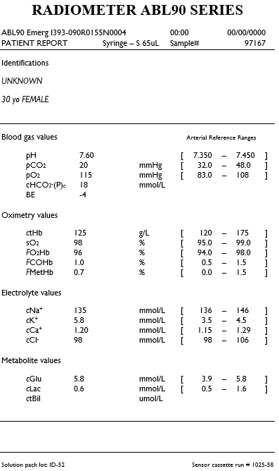
30 year old female
Brought by ambulance with confusion and complaining of ringing in her ears. Her boyfriends report they had an argument earlier today and denies any physical assault.
HR 112, BP 99/62, RR 34, SpO2 100% RA, Temp 37.8, GCS 14 (confused)
What is going on here?
This is an arterial gas on arrival. Albumin is pending…
There is a moderate alkaemia. With a low pCO2 this suggests that the primary pathology is a respiratory alkalosis.
We can then look for secondary acid-base disturbances. Using the 1,2,4,5 rule for metabolic compensation we know that for every 10 of CO2 below 40 the bicarb should decrease by 2 in acute cases and 5 in chronic cases.
Therefore in this case, the expected bicarb is 24 - 2 x 2 = 20 if acute or 24 – 2 x 5 = 14 if chronic. Chronic respiratory alkalosis is not that common. Causes include third trimester pregnancy, high altitude and chronic liver disease. There is nothing to suggest chronicity in the stem and therefore with a bicarb that is slightly lower than expected and a negative base excess there is also a metabolic acidosis present.
To look at the metabolic component using Fencl-Stewart
SID 135+6-98 = 43 (within +/- 2 of 42 so no real chloride effect)
Albumin effect unknown so assumed to be normal
Normal lactate
Therefore, there is a mild unexplained acidosis with a base excess of -4
Using a traditional approach
AG = 135-98-18 = 19 so there is a mild HAGMA. Causes – recall LTKR. Is this a toxin, ketones or renal?
Delta ratio = 19-12/24-18 = 7/6 so this is a pure HAGMA
Other information from this gas
- Mild to moderate hyperkalaemia. Expected K for this pH = 4.0
- AA gradient can be calculated: 150 – 1.25 x 20 -115 = 10 (normal).
Clinical interpretation. The answer is in the stem. This presentation is typical for a salicylate overdose. The pattern on the gas of a combined respiratory alkalosis and metabolic acidosis is consistent with this.