
10 June 2020 - Geriatrics
10am-11am – Richard – Delirium
11am-12pm – Ken – Acute assessment of the older person and frailty
12pm- 1pm – lunch and ED stuff (recap from last week, ECG, ABG and image of the week)
1pm-2pm – Min – Geriatric Medicine and community services at St Vincent’s
1pm-3pm - OSCE Group - Brendan
Key Learning Points
Delirium
Hypoactive delirium has worse outcomes than other forms of delirium
Don’t forget the importance of non-pharmacological treatments of delirium as first line therapies.
Reassurance, orientation techniques, avoid restraints, hydration and nutrition, noise reduction, familiarity (Get out of the ED or ICU)
If pharmacological therapy is needed, use the lowest dose of antipsychotic
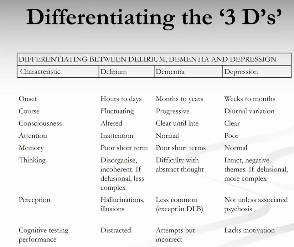
Differentiating between delirium and dementia is one of few geriatric topics easily examinable in ACEM exams. And hence it comes up a lot.
Frailty
Associated with increased risk of falls, worsening mobility/disability, higher hospitalization rates and death
Frail people need more medications resulting higher adverse drug reaction events.
Gait speed is a measure of health and is reflective of whole body function (brain, CVS, respiratory etc). Fast walkers overall have a greater life expectancy
ECG of the Week
55 year old male presents with a gradual worsening of dyspnoea over the past month. He has a history of a lobectomy for lung Ca two years ago. No chest pain.
This ECG is a spot diagnosis and suggests a massive pericardial effusion. Key findings are sinus tachycardia, electrical alternans and small QRS voltages. The next urgent test is a bedside echocardiogram to confirm the diagnosis.
Electrical alternans is caused by a pendular heart movement within the pericardial fluid. Therefore the effusion has to be large/massive suggesting non-acute onset. These patients often do not need emergency pericardiocentesis and can be managed by cardiothoracics. In an acute tamponade, such as from trauma or ruptured vessel we will not see these ECG changes as tamponade occurs with a small volume of fluid/clot contained within the fibrous pericardium.
Image of the Week
This patient was brought by ambulance after falling from a motorbike at 20kmph. The bike fell onto his L ankle with a ? open fracture.
O/E. Laceration to lateral ankle with tenderness & deformity over the wound, severe pain on passive ankle movement.
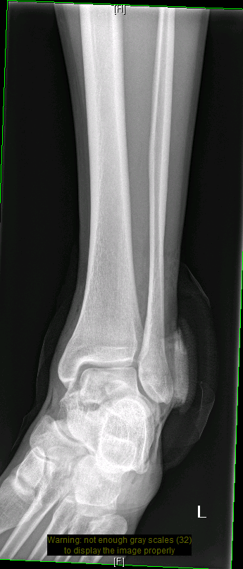
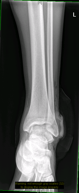
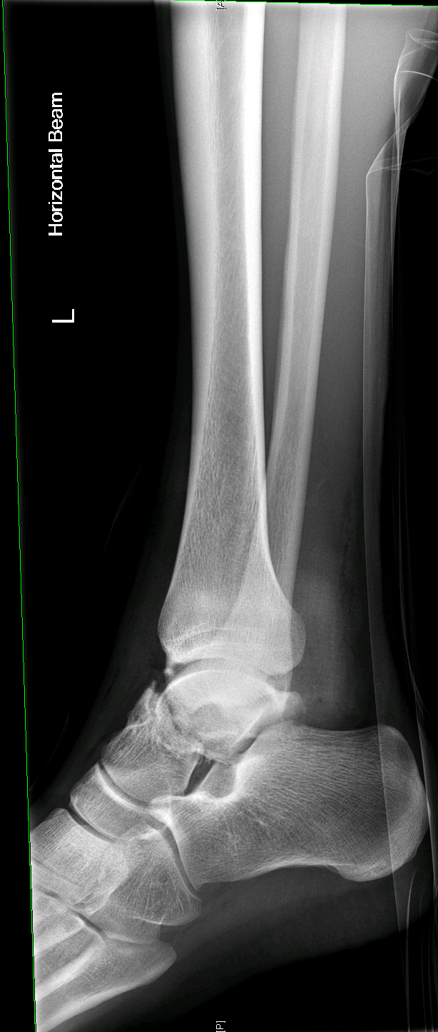
The X-rays show a talar neck fracture with subtalar dislocation. There is gas in the soft tissues suggesting an open fracture. Reduction in the ED is required followed by prompt orthopaedic management. These injuries give a risk of avascular necrosis and the Hawkins classification can be used to estimate this risk.
Interesting Blood Gas
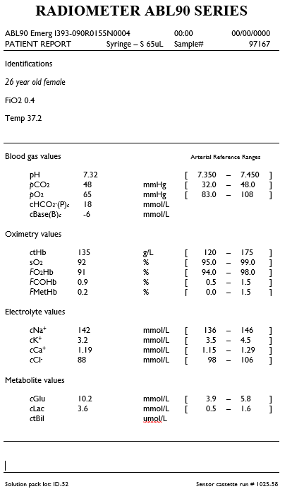
A 26 year old asthmatic with a history of ICU admissions presents with acute respiratory distress. She has been treated by her GP for 2 days with bronchodilators and glucocorticoids.
Vital signs are:
HR 120, BP 110/70, RR 30, SpO2 92% with FiO2 40%.
The respiratory registrar is keen and takes an ABG as shown. What information can you gather from it?
The patient’s albumin was normal.
Interpretation
There is a mild academia with low bicarb and high pCO2 suggesting a primary mixed respiratory and metabolic acidosis.
Using a traditional approach:
AG = 142 - 88 - 18 = 36
Delta Ratio: (36-12)/(24-18) = 24/6 = 4!
This suggests a HAGMA and a coexisting metabolic alkalosis
Using Fencl-stewart
Measured base excess is -6
SID = 142+ 3.2 - 88 = 57. Therefore chloride effect on BE is +15. I.e. there is a hypochloraemic metabolic alkalosis
No albumin effect
Lactate effect is -2.5
The sum of these effects is 12.5, with the measured base excess -6 therefore there is an unexplained - 18.5 suggesting another unexplained metabolic acidosis.
This doesn’t really make sense that there is such a significant unknown metabolic acidosis unless this patient is also in DKA? Or has also drunk ethylene glycol??
This particular blood gas was taken from a text book that focuses on the traditional method only and I suspect that the chloride value was “made up” rather than real physiology.
So summary of the acid-base status is that there is a triple disturbance of a metabolic acidosis, metabolic alkalosis and respiratory acidosis.
Causes of the metabolic acidosis could be lactate from salbutamol use. Ketones unknown. Toxins unknown. Kidney function unknown.
Cause of the metabolic alkalosis is unknown. Remember the acronym GROE. See - previous post on gas interpretation. Has this patient been vomiting? Is there renal dysfunction? Is the patient cushingoeid from long term steroid use?
Other information from the gas
Oxygenation. The gas shows critical hypoxia with a pO2 of 65 with a patient on oxygen therapy. We can calculate an Aa gradient. = 300 - 1.25 x 48 - 65
= 175.
Therefore raised Aa Gradient suggesting VQ mismatch.
There is also a mild hypokalaemia.
Overall summary
To see these changes on a blood gas suggests critical, life-threatening asthma. The metabolic component can’t be completely explained but any acidosis in an asthmatic suggests severe illness.