
24 June 2020 - Paediatrics
Program
1000-1015hr – Welcome and log in to Microsoft teams, Trivia!
1015-1030hr – Recap of last week’s Paeds Regional Teaching
1030-1100hr – Seizures in Paediatrics – Ben Lee
1100-1130hr – Fever and Sepsis in Paediatrics – Max Grant
1130-1140hr – coffee break
1140-1230hr – APLS Quiz – Brendan
1230-1250hr – ECG/gas/image of the week – Alex/Mark/Matt
1250-1305 – lunch break
1305-1310hr – 5 min Tox – Paed theme - Jacqueline
1310-1400hr – SAQs - Jacqueline
1330-1500hr – Ultrasound group with Mark –please arrange direct, first dibs to those working at 3pm
Key Learning Points
Paediatric Seizures
A simple febrile seizure has all of the following:
Fever
Generalised tonic-clonic seizure
Duration < 10minutes
Complete recovery within 1 hour
No recurrence within the same febrile illness
A complex febrile seizure is everything else (i.e. focal seizure, > 10 minutes, not recovered within 1 hour, recurrence)
Note there is the concept of an afebrile febrile seizure. These occur in an acute infectious illness, particularly gastroenteritis, without a documented features but a seizure otherwise consistent with a simple febrile seizure
WETFLAG is a common acronym that can be used as a memory aid for preparing to receive a sick child
W: Weight: 2 x (Age + 4)
E: Energy: 4J/kg
T: Tube: ETT: Age/4 + 4 (uncuffed tube - lower size for cuffed tube)
F: Fluids: 10-20mls/kg
L: Lorazepam (more likely to use midazolam 0.15mg/kg)
A: Adrenaline: 10mcg/kg
G: Glucose: 2mls/kg of 10% dextrose
Recent studies have shown no difference in efficacy between valproate, levetiracetam, phenytoin as second line agents (but levetiracetam is often easier to administer for unfamiliar staff)
We discussed the algorithm to managing status epilepticus in children a few weeks ago. Click here.
Fever and Sepsis in Children
To treat or not treat a fever? There are lots of approaches. Generally speaking, treat the child that is miserable or in pain (i.e. sore throat) rather than the temperature. In the ED, treating the fever often allows a better examination, a happier child and more satisfied parents. Often, a child that has been brought to the ED will warrant a dose of paracetamol/ibuprofen.
Sepsis Kills. RCH has a great clinical practice guideline that was recently updated. Check it out here.
Key principles in managing severe sepsis or septic shock
Early recognition/seeking senior help
Assess airway and breathing and administer oxygen if required
Rapid vascular access
Empiric antibiotic therapy
Carefully titrated fluid resuscitation
Early initiation of inotropes
Early involvement of critical care services
Source control
Frequent reassessment
APLS Quiz
All of the APLS algorithms are freely available. You can download the APP onto your smartphone for easy access when the sick child suddenly appears in the ED

ECG of the Week
Thanks Kimson for the case.
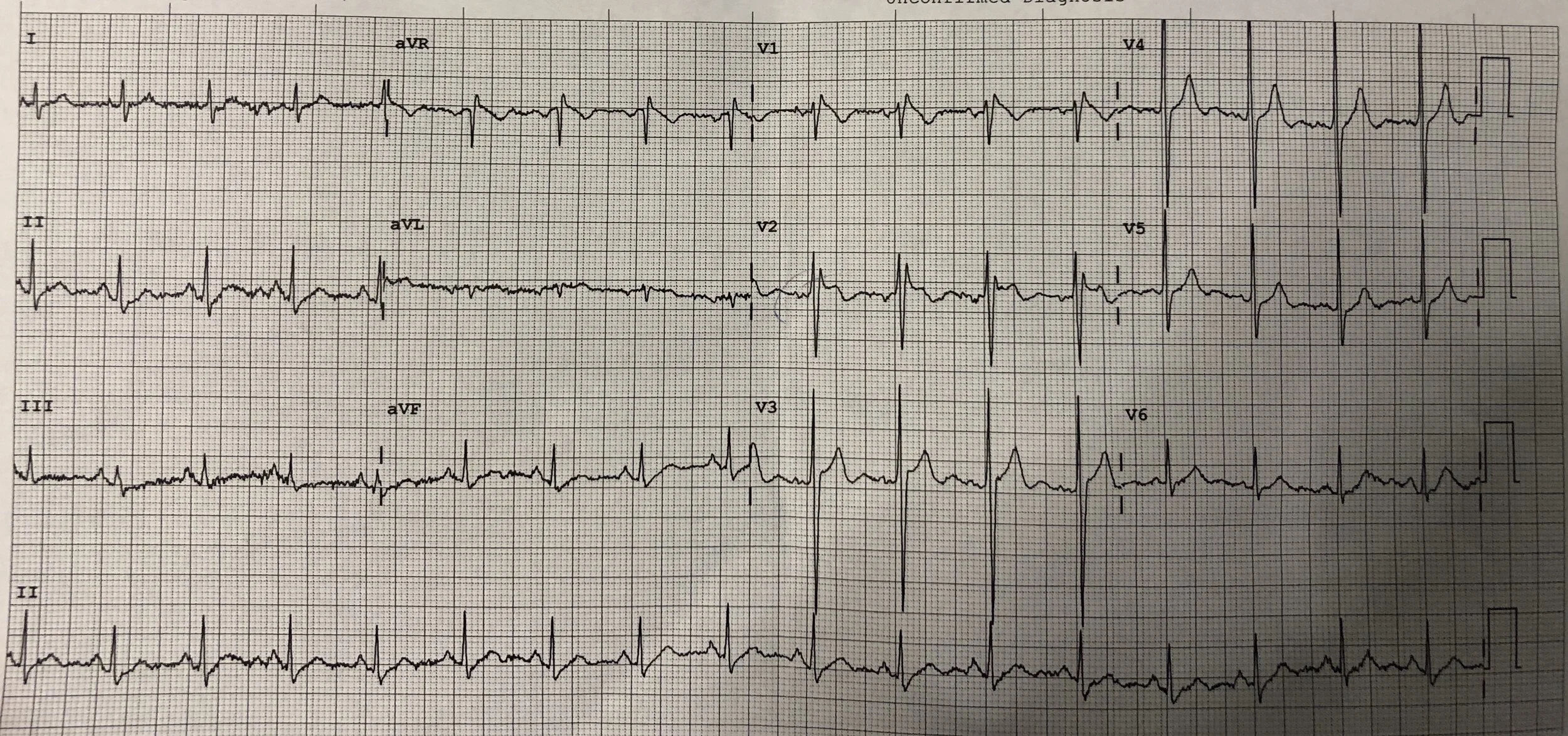
A 60 yo male presented to the ED with pneumonia. He was febrile at 40 degrees but not hypoxic and able to be discharged home on oral antibiotics. A question was raised about the possibility of Brugada Sign.
There are three repolarisation patterns associated with Brugada syndrome when found in more than one right precordial leads (V1 to V3):
Type 1: It is characterised by a prominent coved ST-segment elevation displaying J-point amplitude or ST-segment elevation ≥2 mm, followed by a negative T wave.
Type 2: It has ≥2 mm J-point elevation, ≥1 mm ST-segment elevation and a saddleback appearance, followed by a positive or biphasic T-wave.
Type 3: It has either a saddleback or coved appearance, but with an ST-segment elevation <1 mm.
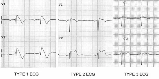
It should be stressed that type 1 is the only ECG diagnostic pattern of Brugada Syndrome, while types 2 and 3 should only be considered suggestive of the disease.
Brugada Sign (the ECG findings) without clinical features is of unknown significance.
Brugada Syndrome is considered when there is Brugada Sign and one of the following clinical features:
Documented ventricular fibrillation (VF) or polymorphic ventricular tachycardia (VT).
Family history of sudden cardiac death at <45 years old .
ECG Type 1 - Coved-type ECGs in family members.
Inducibility of VT with programmed electrical stimulation .
Syncope (and seizures?).
Nocturnal agonal respiration.
In the above case, the ECG does not fit the criteria for Type 1 Brugada Sign. There is an incomplete RBBB and at the J point in V1 there is <2mm ST elevation. The patient did not have any of the clinical features of Brugada Syndrome. A subsequent ECG one hour later showed improvement of these findings. We know that fever can unmask Brugada Sign on ECG and the patient was referred to cardiology outpatients for further opinion.
Here is the link to the article by Professor Brugada
Image of the Week
A 76 yo female fell on her outstretched hand. Her X-rays are included below.
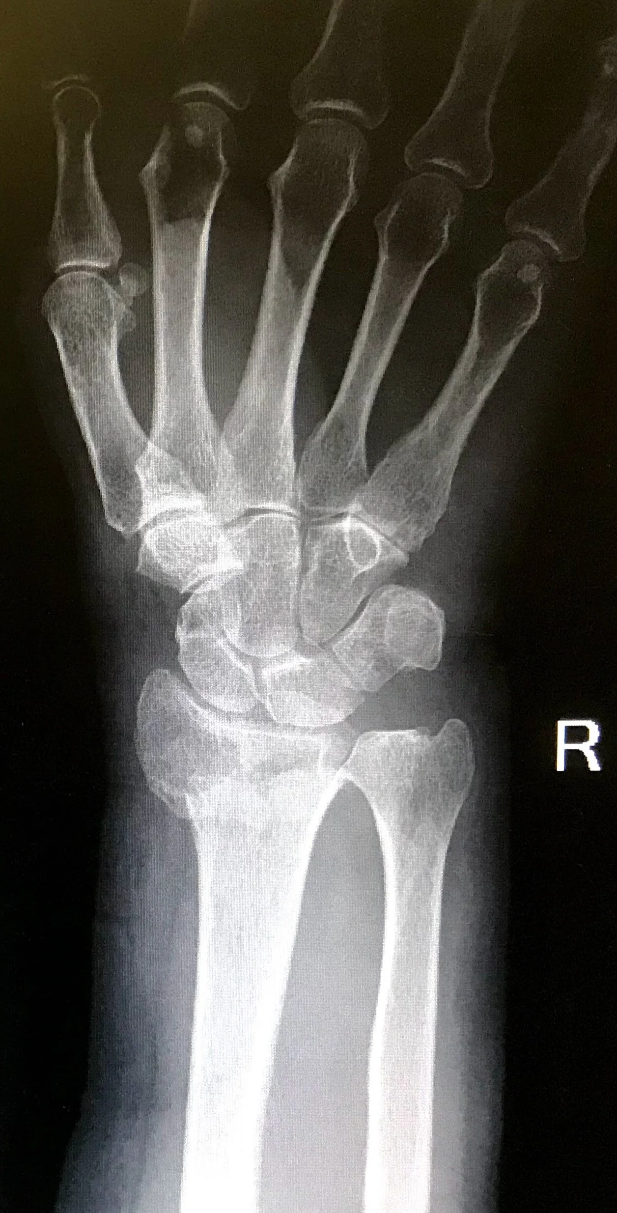
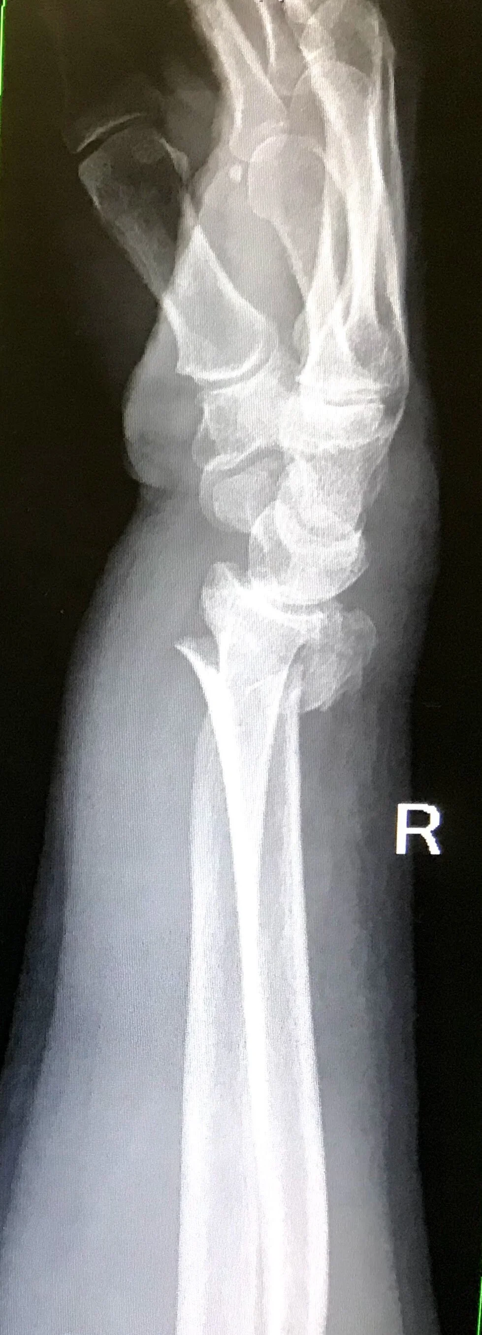
Right comminuted intra-articular distal radius
These xrays show a distal radius fracture. It is impacted with positive ulna variance, radial shortening, loss of radial height & radial inclination and dorsal angulation.
How do you know which of these need ED reduction if it is not obvious? This hospital has a distal radius fracture pathway available on the intranet. In summary fractures need reduction if any of the following are present:
Neurovascular compromise
Dorsal tilt > 10 degrees
Radial shortening > 5mm
Radial inclination < 15 degrees
If unsure, give the orthopaedic registrar a call.
Interesting Blood Gas
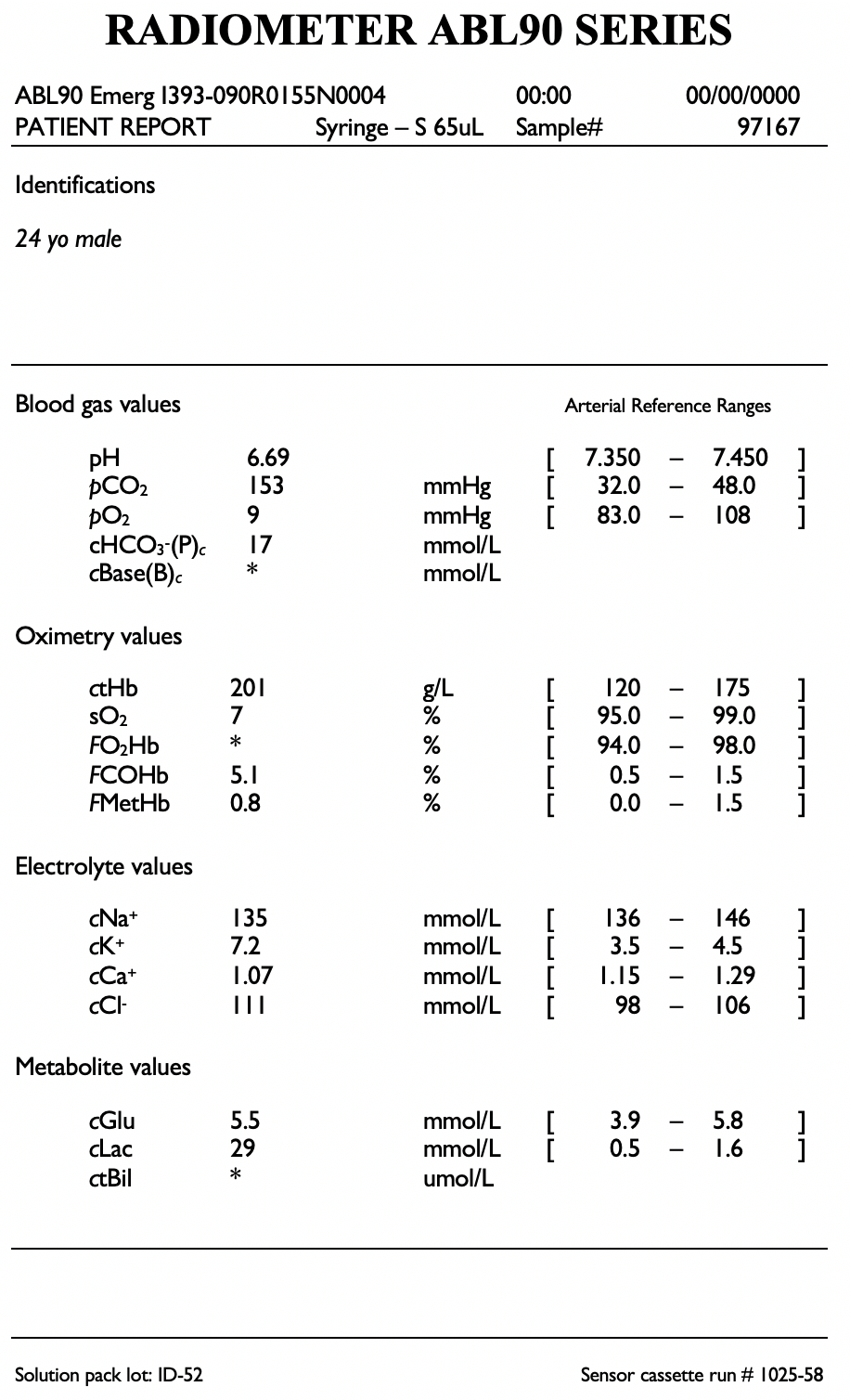
A 24 yo male with a history of schizophrenia presents with priapism of 4 hours duration. A cavernosal gas is taken and shown.
A blood gas can help differentiate between ischaemia (low flow) priapism and non-ischaemic (high flow) priapism if it is not evident in the history.
If the pH > 7.25, pO2 > 30 or pCO2 < 60 then considered non-ischaemic.
This gas therefore is suggestive of ischaemic priapism and urgent management is required.