
23 September 2020 - Trauma
Program
10-1015hr – Welcome back, feedback on Regional teaching days
1015-1030hr – EDNAV is here, GTG! - Jacqueline
1030-1130hr – Shoulder Dislocation – Neil Cunningham - *** Look at his website for pre-reading*** Link below.
1130-1145hr – break
1145-1215hr – ECG of the Week, Image of the Week , Gas of the Week - Alex, Maria, Mark
1215 –1245hr – Spinal injuries – Chris Loo
1245-1315hr – Chest trauma in the elderly – Stuart Howard
1315-1330hr – break
1330-1445hr – SIM session for Maria, Jess and Jonathon – advanced ALS
1330-1400hr SAQs – Jacqueline
1330hr-1500hr – Ultrasound: 1:1 Mark
Key Learning Points
Shoulder dislocations
Have you visited Neil’s dislocation website yet? Check it out. https://dislocation.com.au
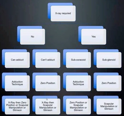
Neil’s suggested algorithm for managing anterior shoulder dislocations
Care of the patient with a shoulder dislocation should be individualised for that patient based on the type of dislocation present (i.e subglenoid or subcoracoid or other), the position of the shoulder (i.e. adducted or not) and patient factors (i.e. severe pain)
Always consider your plan B if your first attempt does not work. Declare this to the patient at the beginning
Get an xray first if you suspect a possible humeral shaft injury - i.e. a larger force injury
Look at shoulder reduction techniques on Neil’s website above
Learn multiple techniques so that you can comfortable move between techniques if the first is not successful
Spinal Injuries
Spinal cord lesions above C5 usually require intubation an mechanical ventilation (often commenced pre-hospital)
Neurogenic shock can result when there is a spinal cord injury above T6. It is a form of distributive shock as there is a loss of sympathetic vasomotor tone resulting in vasodilation, reduced venous return and hypotension. The patient is often bradycardic.
Spinal shock is a reversible spinal cord dysfunction. It can be viewed as a ‘concussion’ of the spinal cord and characterised by temporary loss of neurologic function and autonomic tone below the lesion. In the ED, this is not a diagnosis that we can make as it can last for 72 hours. It can also produce hypotension and bradycardia.
In the hypotensive trauma patient, the hypotension is much more likely to be from haemorrhage or obstructive shock. Neurogenic shock is a diagnosis of exclusion. The treatment is vasopressors.
CT cervical spine is the standard of care for clearing for injuries. Cervical spine X-rays are simply not good enough.
Sensitivity of X-rays for thoracolumbar fractures are poor. Usually 60-65% sensitivity. Get a CT!
Patterns of injury:
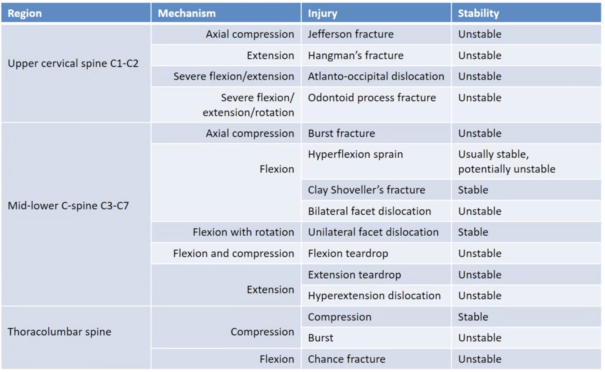
Transverse process fractures are commonly seen in the thoracolumbar spine and are considered stable
Chest trauma in the elderly
Did you know that St Vincent’s has a guideline for the blunt chest trauma in the elderly? It’s on the intranet.
Low mechanism trauma does not mean low-significance injuries in the elderly
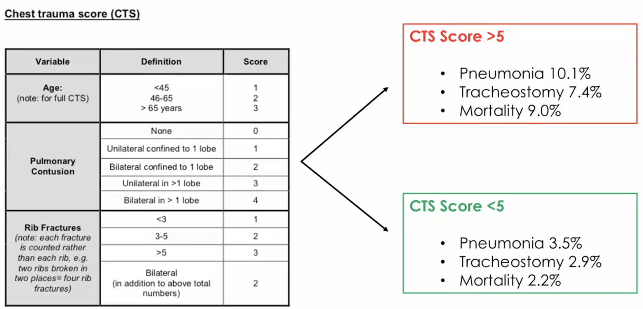
Patterns of blunt chest injuries
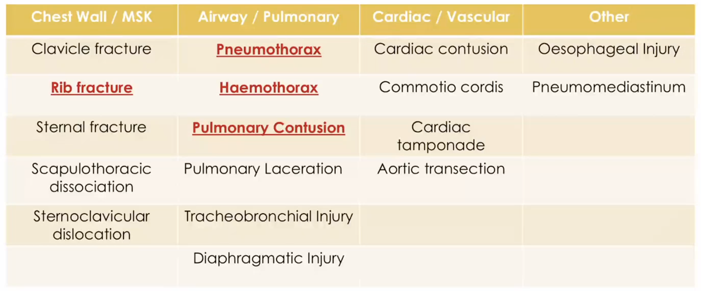
In the elderly, for every successive rib fracture the mortality increases by 18% and risk of pneumonia by 27%.
When considering specific complications of rib fractures:
Ribs 1-3: vascular injuries, cardiac trauma
Ribs 4-9: pulmonary/pleural injury
Ribs 9-11: intrabdominal pathology
CXR will miss 50% of fractures. Have a low threshold in the elderly to order a CT chest after the CXR if imaging is required
ECG of the Week
Thanks Luke for the case this week.
A 47 yo male presented during the night shift with 6 hours of palpitations. He has a history of SVT in which he usually performs a valsalva manoeuvre which resolves his symptoms.
Here is his ECG on arrival.
He is alert, BP 110/80.
Remember my approach to all arrhythmias. This is particularly helpful when the ECG looks bizzarre
Is it fast or slow?
Is it broad or narrow?
Is it regular or irregular?
This ECG is fast, broad and irregular. There are only a few diagnoses that can cause this combination. These include:
AF with WPW
AF with a BBB
AF with another reason for broad QRS complexes such as hyperkalaemia or TCA overdose
A Flutter with a variable block and one of the above (stretching it here)
This particular ECG also shows a rate of 180bpm with periods that appear > 200 bpm. There is also run of non-sustained VT (beats 6-10). Of particular note, the QRS complexes are of variable morphology. This fits with the diagnoses of AF with WPW.
Pearls
Think AF with WPW in any:
Irregular broad complex rhythms with rates > 200
When the QRS complexes vary and look ‘bizarre’ or ‘complex’
These patients should be cardioverted. Procainamide is the only universally safe medication for chemical cardioversion. Our ICU has it, but not many of us have ever used it.
Do not give AV blocking drugs. Adenosine, CCBs and possible amiodarone can precipitate a cardiac arrest with VF and VT.
Image of the week
A 55 year old man is brought in by ambulance following a heroin overdose. He received bystander CPR at the scene and was given IM naloxone by paramedics. His SpO2 is 80% on RA and he is complaining of chest pain and shortness of breath. A CT chest with contrast is performed.
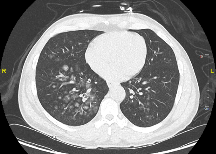
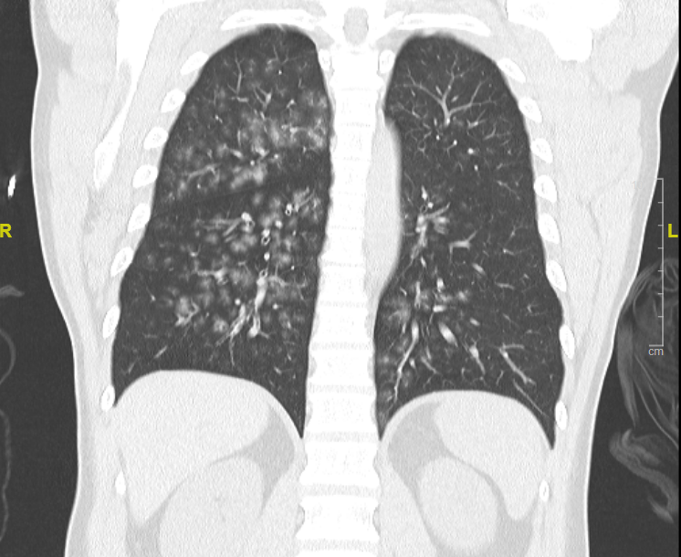
These images show ground glass opacification of which there are many causes.
Unlike consolidation where the pulmonary vessels are obscured, there is no obscuration of these vessels in ground glass changes.
Most commonly it is caused by the alveoli filling with fluid (pus, oedema, blood, exudate, tumour). In this patient’s case, it was aspirated vomitus.
Broadly speaking the causes of ground glass changes can be classified as
infectious processes
chronic interstitial diseases
acute alveolar diseases
other causes
Check out the article on radiopaedia for more information.
https://radiopaedia.org/articles/ground-glass-opacification-3
Gas of the Week
We ran out of time to discuss the gas from the last teaching session. Please go back and have a look through.