
30 September 2020 - Gastroenterology
10-1015hr – Welcome and recap Camilo
1015-1045hr – GI bleed – Jess
1045-1100hr – Discussion of massive GIB case – Joel
1100-1130hr - Inflammatory Bowel Disease and Extra-abdominal manifestations – Hope
1130-1200hr – Journal Club – paper below– Ben and Hamed
1200-1215hr – break
1215-1245hr – ECG/image/gas of the week – Alex, Maria, Mark
1245-1315hr – interesting but sad case – Ben
1315-1400hr – Gastro SAQs – Sarah
1315 - 1430hr – SIM session with Alex and Brendan – Luke and Matt
Journal Club
Brown SGA, Ball EL, Perrin K, et al. Conservative versus Interventional Treatment for Spontaneous Pneumothorax. The New England journal of medicine. 2020; 382(5):405-415. PMID: 31995686 [full text]
This paper has been discussed broadly in the FOAM world this year.
Key learning points
GI Bleeding
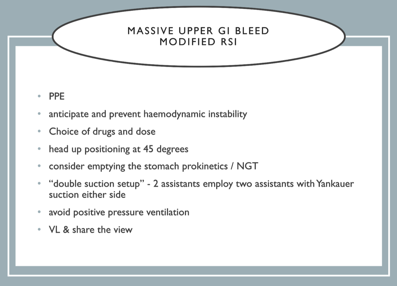
General resuscitative management of the patient with massive GI haemorrhage
Large bore IV access
Fluid resuscitation - preferably blood
Massive transfusion protocol 1:1:1 ratio
Avoid hypothermia, acidosis and treat coagulopathy
Balloon tamponade if pre-arrest/ongoing bleeding as a temporizing measure
? TXA - recent HALT-IT trial showed no benefit of TXA with known adverse effects
Pantoprazole 80mg bolus
Definitive care - endoscopy
Adjunctive treatment for variceal bleeds
Octreotide 50mcg bolus then 50mcg/hr infusion (alternative is terlipressin)
Ceftriaxone 2g IV
ECG of the Week
A 65 yo male was brought by ambulance. Signal one pre-notification with the below ECG taken by paramedics. HR 25 with no recordable blood pressure.
Complex PMHX including mild CCF, paroxysmal AF, ESKD on dialysis, bilateral PEs, T2DM, TB peritonitis.
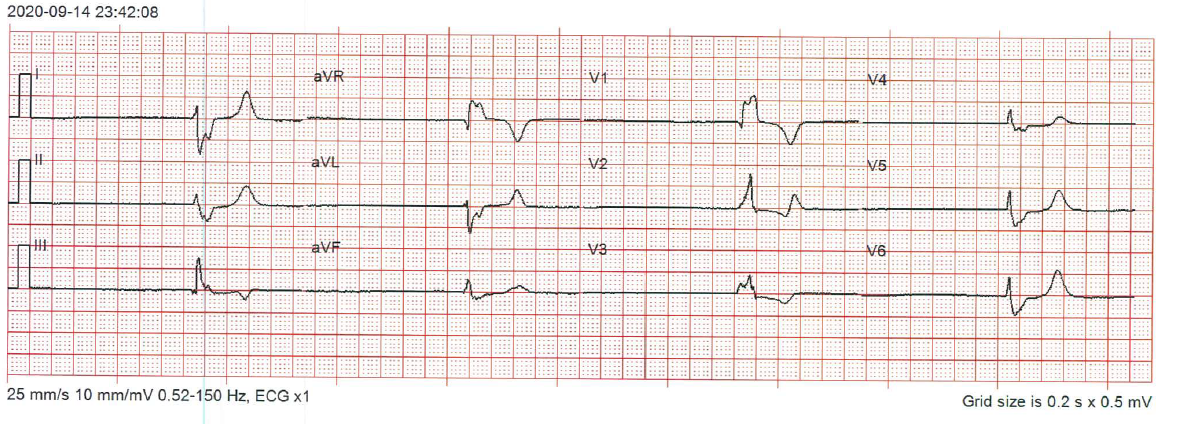
This ECG show a bradycardia of about 24 beats per minute. It appears regular but it is difficult to tell when the rhythm is so slow. There are no discernable P waves. The QRS complexes are very broad (about 200ms) and has a RBBB appearance.
Differential diagnoses for this patient include hyperkalaemia, ischaemia and toxicity from BB/CCB/Digoxin.
As the patient arrived with AV the BP was 90/60 post a fluid bolus.
The patient’s K was 6.5 on blood gas. Although not markedly high, the patient’s baseline K was in the normal range. In the absence of other clear reasons, it was felt that this was a major contributor in the cause of the arrhythmia.
Hyperkalaemia was treated (calcium, insulin, dextrose) and dialysis was organised overnight for the patient.
Image of the Week
24F presenting following a motorcycle injury at low speed. Whilst travelling at 5Km/h, she accidentally swerved and fell off the motorcycle, landing on her left side with the motorcycle landing on her legs. Protective leathers with knee pads worn. She has an isolated left knee injury and is unable to weight-bear. An X ray of her left knee is shown.
The X-ray shows a lipohaemarthrosis, a tibial spine fracture and possibly a segond fracture although this is not clear.
This injury is concerning for serious ligamentous damage and possible ACL repair.
This patient should go on to have a CT.
Gas of the Week
A 36 yo female with a history of alcohol abuse presents with 2L of frank haematemesis. She arrives in the ED with a thready pulse and no recordable blood pressure.
This arterial blood gas was taken about 90 minutes into her resuscitation. A massive transfusion protocol was running.
She was on 15L oxygen via a non-rebreather mask at the time of this gas.
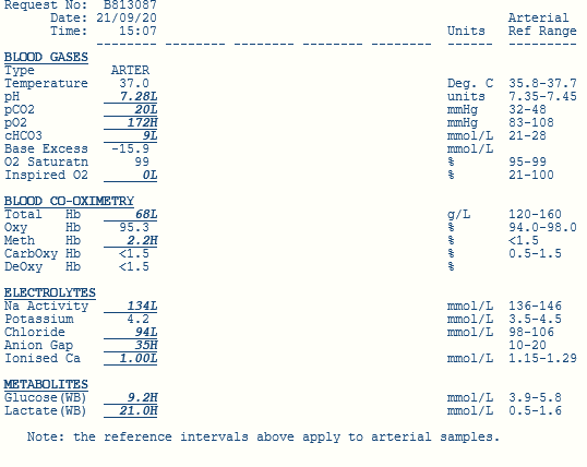
The primary acid-base disturbance is a moderate metabolic acidosis as evidenced by a base excess of -15, low pH and low bicarb.
Using Fencl-Stewart
SID = 134+4-94 = 44. This is within the margin of error for chloride so no significant effect on the base excess
Lactate effect = -20. This change in lactate is likely secondary to hypo perfusion from shock but liver failure might also be a contributing factor.
Albumin effect = unknown. Likely hypoalbuminaemic from liver failure.
Using a traditional approach
AG = 31 or 35 if using the potassium. Therefore there is a HAGMA
Delta ratio = (31-12)/(24-9) = 19/15. Therefore there is an isolated HAGMA - almost certainly due to the high lactate
The expected CO2 using Winter’s formula is 1.5 x 9 + 8 = 21.5. Therefore there is appropriate respiratory compensation.
There is a raised Aa gradient that could be explained by aspiration or pneumonia in this patient.
Other important things to note include a marked anaemia and also hypocalcaemia. This is particularly important given this patient is receiving a massive transfusion and there could be citrate toxicity from receiving blood products. This patient requires parenteral calcium.