
22 April 2020 - Abdominal
Program
1000-1015: Greeting and recap
1015-1100: Jaundice, acute liver failure and LFT interpretation – Ben
1105-1135: Paediatric surgical emergencies – Mark Rugless
1135-1150: Beware constipation! Abdominal pain in the elderly – Sarah Ward
1200-1210: ECG of the week – Alex
1210-1300: SAQ’s with Brendan and Sarah
Key Learning Points
Jaundice/Liver Failure/LFTs
ALT and AST are transaminases and often reflect a hepatocellular pattern of LFT abnormality
> 10 x the normal upper limit of ALT indicates severe dysfunction
ALT > 1000U/L usually signifies toxins, ischaemia or acute viral hepatitis (A or E)
ALP and GGT elevation together, usually signify a cholestatic pattern of LFT abnormality
Fulminant liver failure has many causes. Ben recommended an acronym DAVES. Drugs, Alcohol, Viruses, Extras (which list all the other causes) and Sepsis
Other pathology abnormalities in liver failure include:
Low albumin
Low clotting factors (except vWF) leading to a raised PT/INR/APTT
Hypoglycaemia
Hyponatraemia
High ammonia
Testicular torsion
Testicles are almost 100% salvageable if operated on within 8 hours, 50% up to 12 hours and 10% up to 24 hours
Do not attempt to detort the testicle in the ED. We can’t reliably tell which way the testicle has torted and as such risk making the situation worse
Ultrasound has a limited utility as 25% of patients with torsion have a normal ultrasound
Elderly constipation
Beware! Serious diagnoses are easily missed here. Consider ischaemic bowel and bowel obstruction in all patients and strongly consider imaging these patients
AXRs may be useful in making an early diagnosis while awaiting CT but are poorly sensitive and specific of serious illnes
ECG of the Week
This 77 yo male presented with vague complaints of weakness.
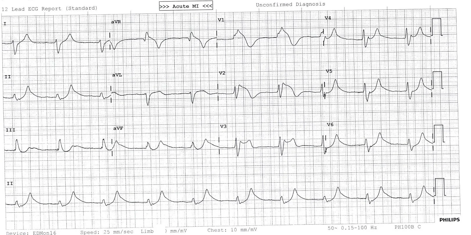
This ECG shows a regular broad complex rhythm with hardly visible P waves if at all. The QRS complexes are broad and bizarre, the T waves peaked.
The patient’s VBG revealed a potassium of 7.7mmol/L
ECG findings with hyperkalaemia are fairly predictable in their progression but do not correlate as well with set potassium concentrations.
In order, these findings typically include:
Repolarisation abnormalities: Peaked T waves
Paralysis of atria: P wave widens and flattens, PR segments lengthens, P waves disappear
Conduction abnormalities: Prolonged QRS with bizarre morphology, any kind go conduction block, sinus bradycardia or slow AF, development of a sine wave appearance
Cardiac arrest: Asystole, VF, or PEA with bizarre wide complex rhythm
Management of hyperkalaemia is expert level knowledge for the FACEM. Initial treatment could include:
Membrane Stabilisation (only give if QRS widening)
10mls of 10% Calcium Gluconate
(Remember Calcium chloride has three times as much calcium per 10mls and probably should only be given via a central line)
Multiple doses may be required
Shift K+ into cells
1mmol/kg NaHCO3 IV - only if patient acidotic
10 units actrapid IV with 50mls of 50% glucose IV
10mg Salbutamol NEB
Increase K+ Elimination
Frusemide (theoretically works but no good evidence)
Dialysis
Resonium - NO. This is not part of the emergency management of the hyperkalaemia patient. It is slow, ineffective and succeeds mainly in constipating patients.
Treat the underling cause
Interesting Pathology
Jacqueline had the following 56 yo patient that presented with jaundice.
His biochemistry is below. Ask yourself:
What could be causing these abnormalities?
What would you look for on examination?
What further investigations would you order?
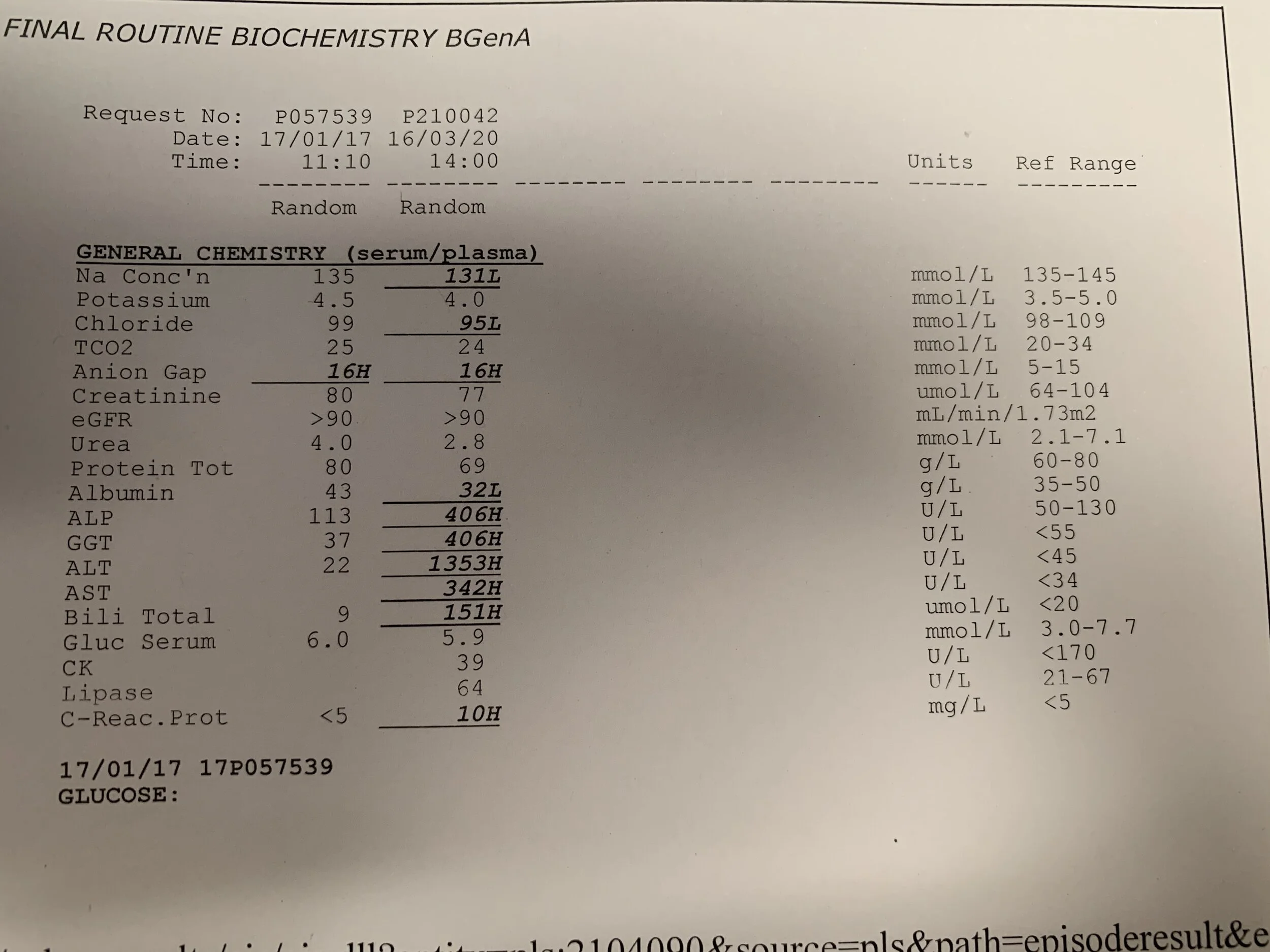
Turns out that this patient had acute hepatitis A on a background of alcohol use and hepatitis C infection.