15 April 2020 - Trauma
Program
1000 - 1015 – Welcome, Recap
1015 - 1045 – Haemorrhage in Trauma – Joel
1045 - 1115 – Torso Injuries – Chest and Abdomen – Jonathon
1115 - 1145 – Epistaxis and Stab wounds to the neck plus ENT competencies – what the college wants you to be able to do – Chris
1145 - 1200 – break
1200 - 1245 – Trauma SAQs – Sarah/Jac
1245 - 1300 – ECG of the week – Alex/Sarah
1300 - 1345 – Anaesthetics Session – update from the COVERT team, Q&A – Ben Slater
1345 - 1400 – Round up, any other issues
Key Learning Points
ENT - Chris Slinger
Chris gave a fantastic talk on ENT emergencies and there were a number of key learning points. Here are the most important ones.
Epistaxis management
Wear PPE!
Simple measures done correctly will stop most cases of Epistaxis.
Pinch the non-bony part of the nose
Apply cotton pledgets soaked in a vasoconstrictor
Give 1g IV transexamic acid
Know how to safely use silver nitrate cautery sticks
Merocel or Rapid Rhino can be used if simple measures fail. Insert all the way in along the floor of the nose
Foley catheters for posterior bleeding does not control the bleed - it simply directs the blood forwards. Definitive treatment is needed.
Nasal fractures
Always look for (and document) the presence or absence of a septal haematoma
If present, these require same day ENT treatment to prevent septal necrosis
ENT Foreign Bodies
This is usually a paediatric presentation
Play can encourage expulsion of the FB from the nose.
A ‘mothers’ kiss is safe and effective more than 50% of the time
Some children require sedation to allow safe removal of the foreign body
Laryngeal foreign bodies should be removed in theatre if at all possible. If not possible, prepare for front of neck access in the event of a complete airway obstruction
Penetrating Neck Trauma
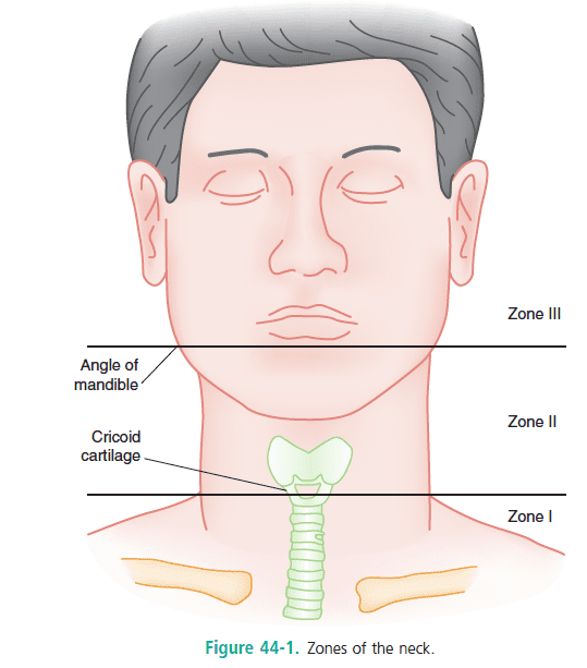
Traditionally there are three anatomical ‘zones’ of the neck that were used to determined whether the patient needs imaging vs theatre exploration. Although this comes up in exams (still) in real practice most of these patients should have CT angiography if stable enough to do so
Any breach of the platysma muscle warrants theatre exploration of the wound. Do not go probing neck wounds in the ED!
There are ‘hard’ and ‘soft’ signs of neck trauma to suggest serious injury requiring theatre. Again, useful for exams but not in practice where imaging and referral are key
Ensure your large bore access is not on the same side as the wound as the subclavian vein may be involved.
Remember that neck wounds can extend into the thorax and mediastinum or into the skull base
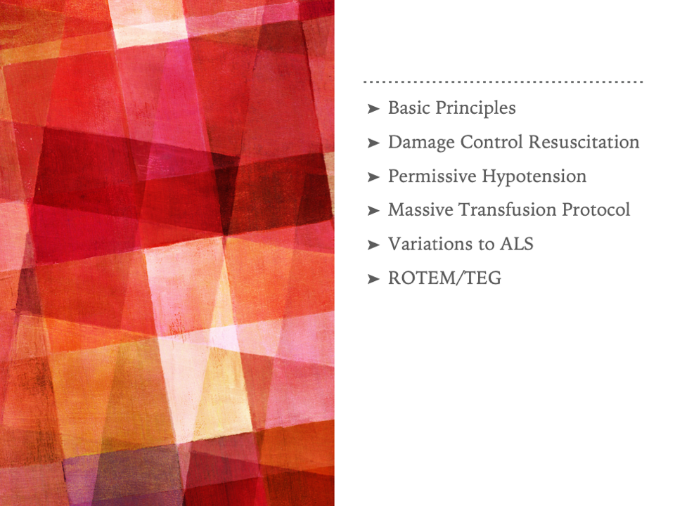
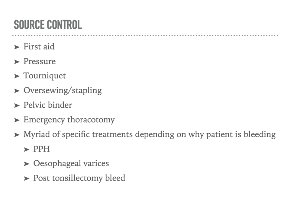
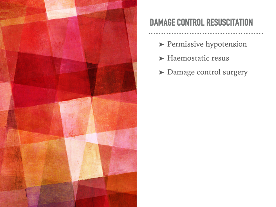
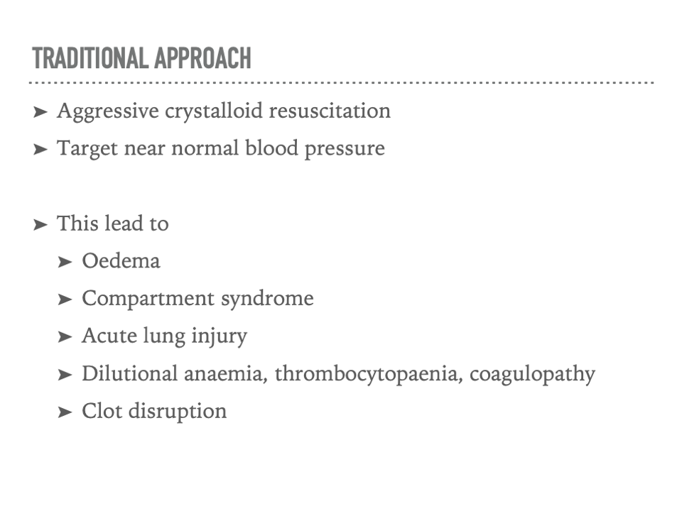
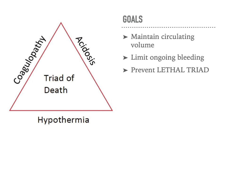
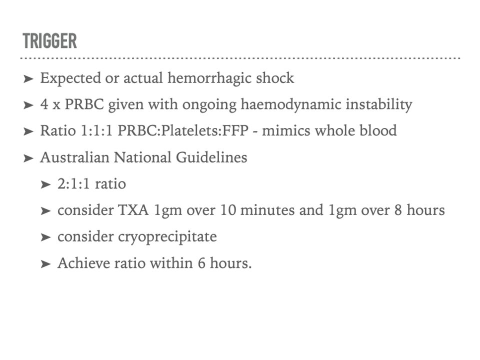
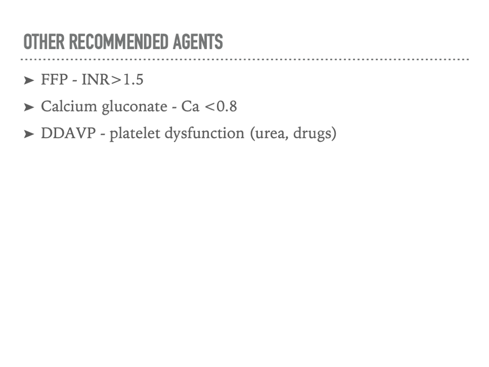
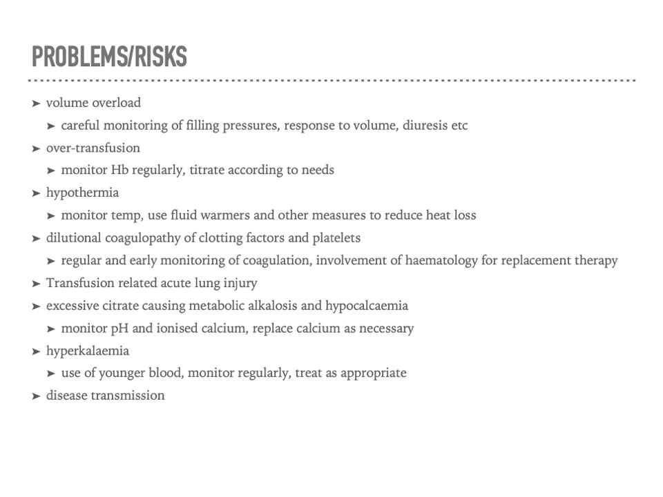
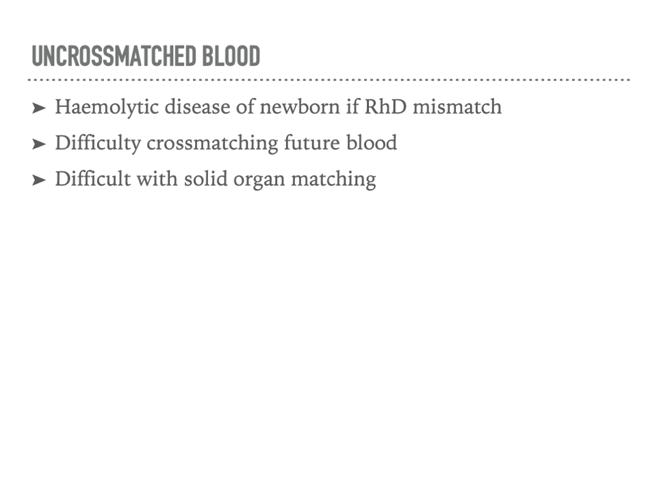
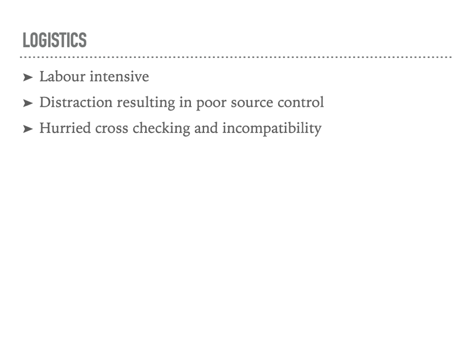
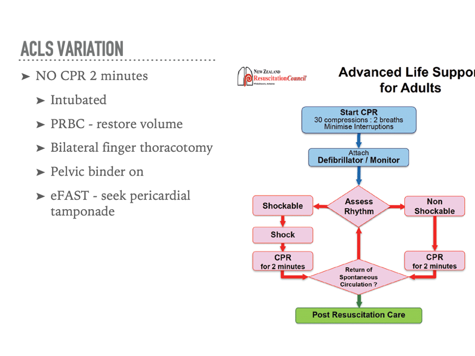
Trauma - Management of Shock - Joel Wilson
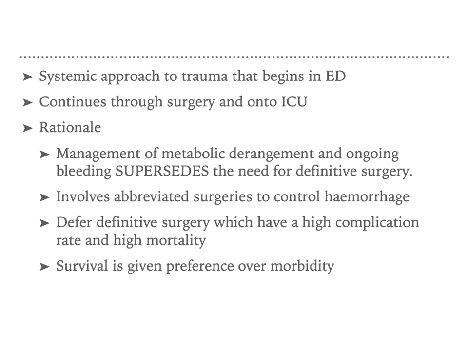
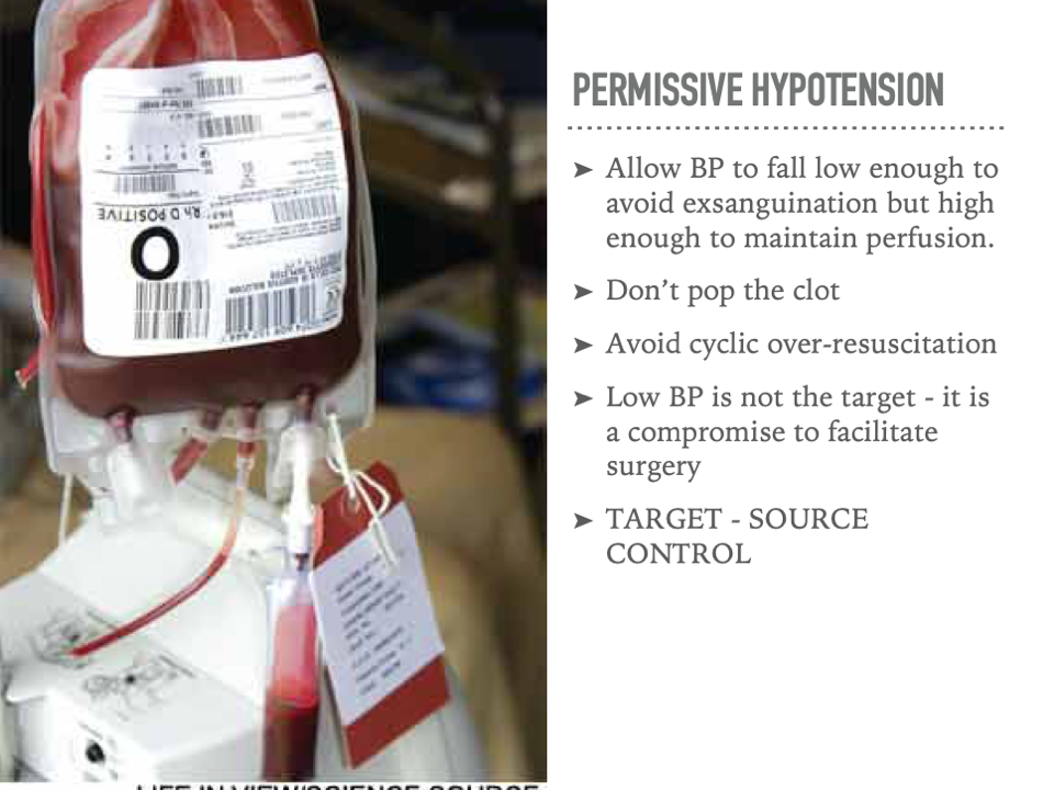
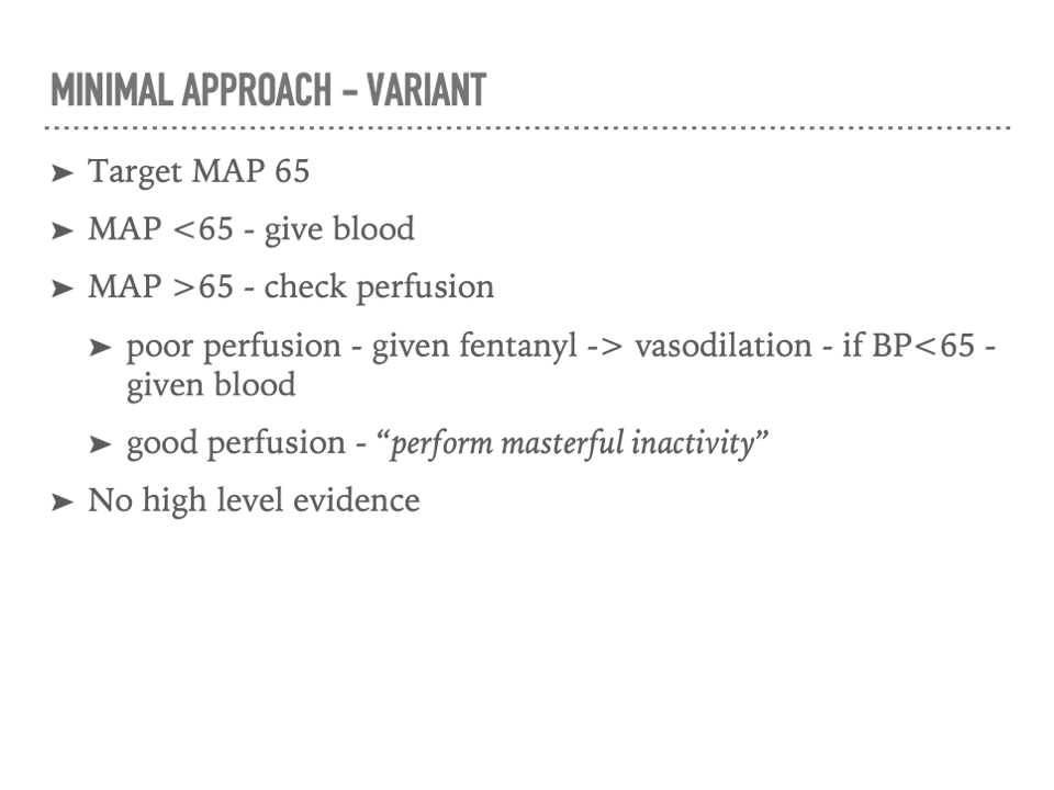
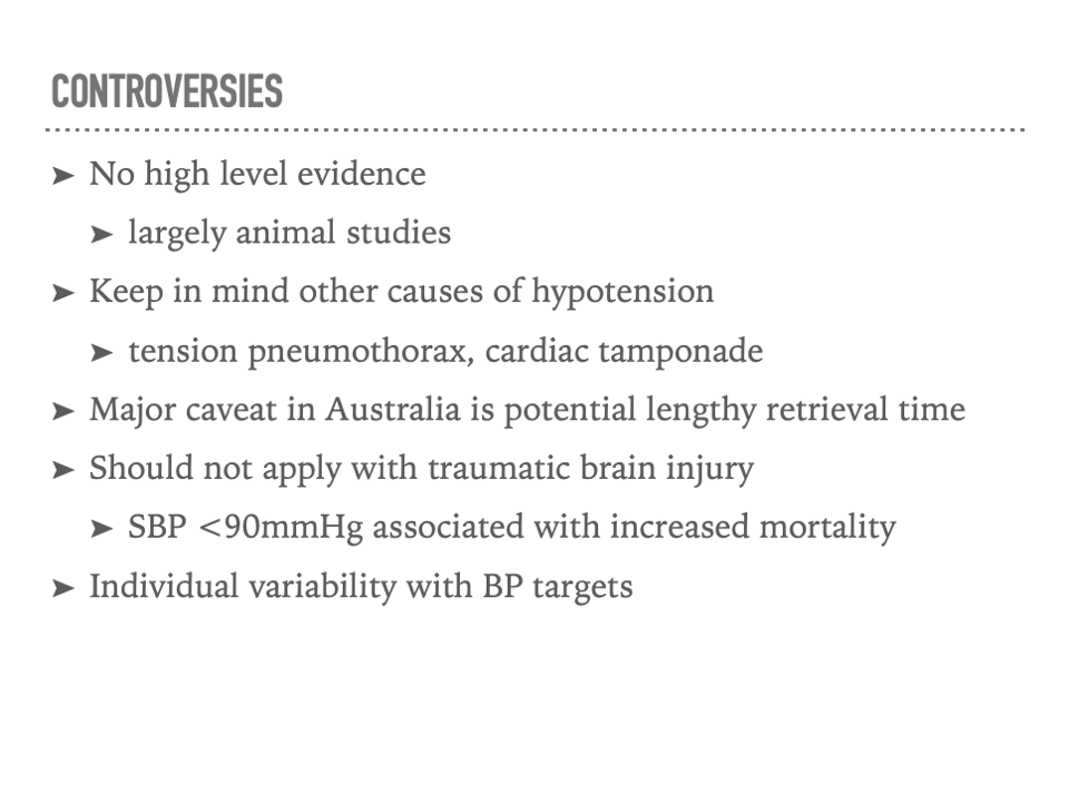
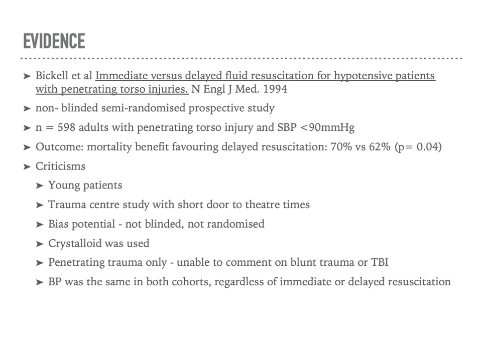
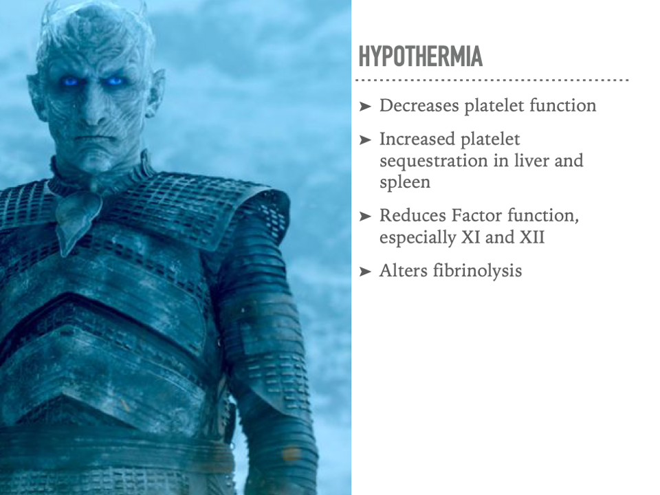
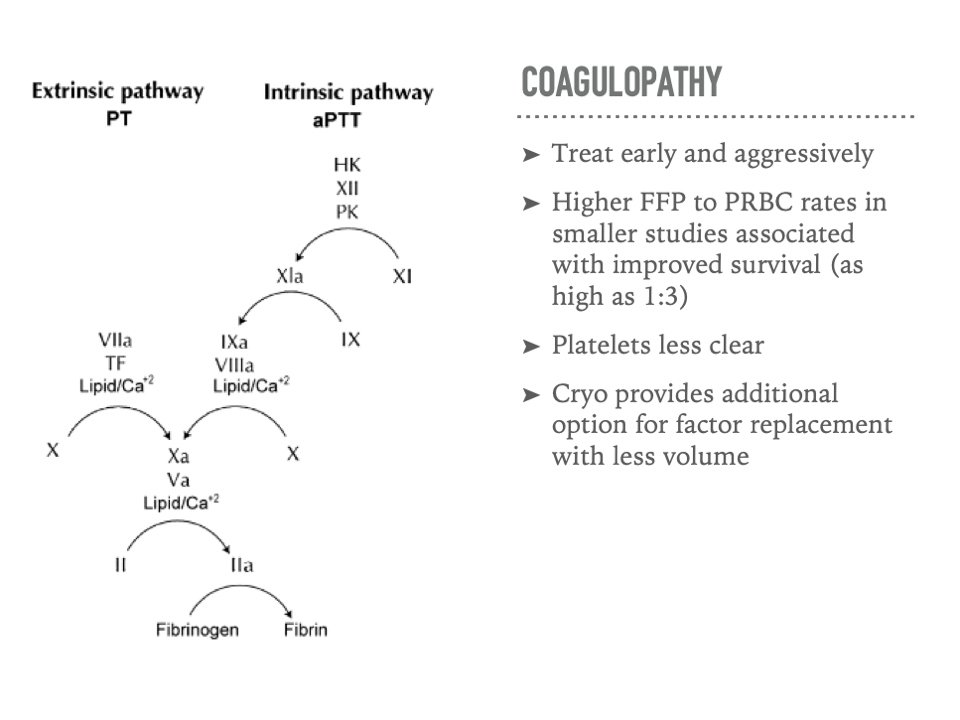
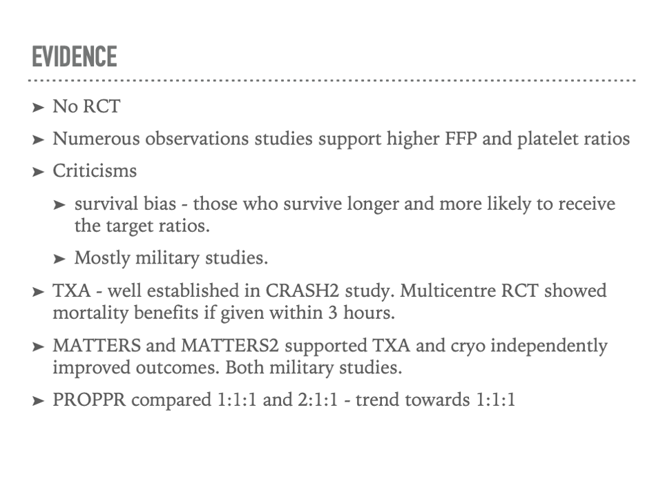
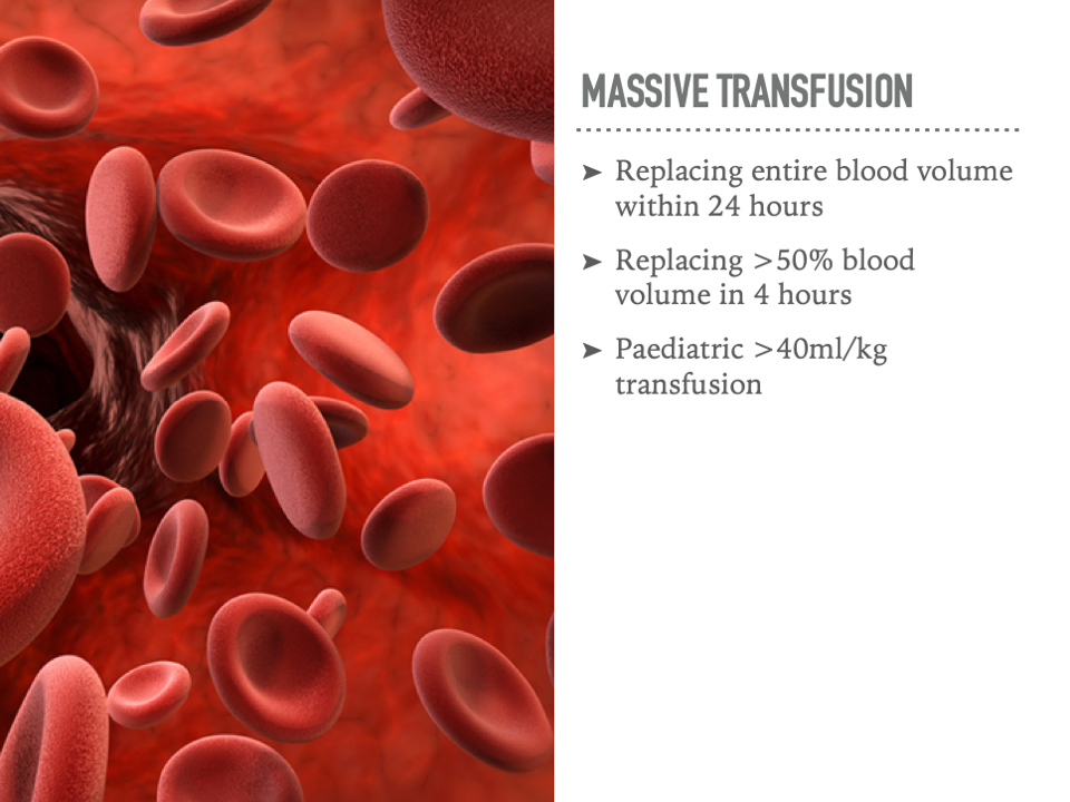
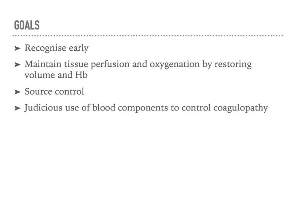
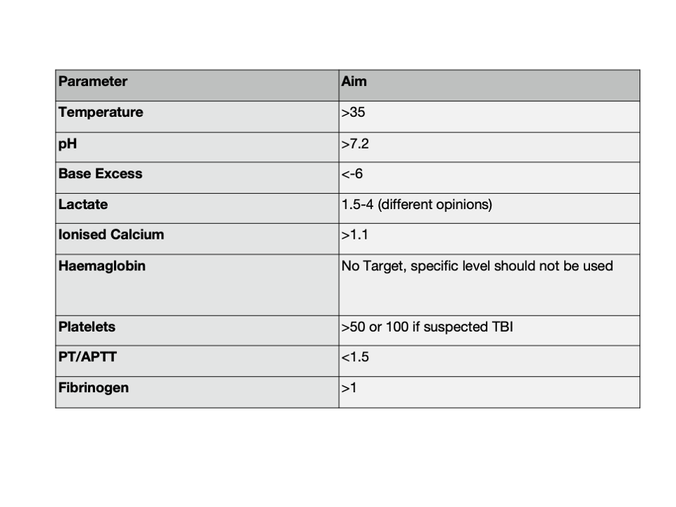
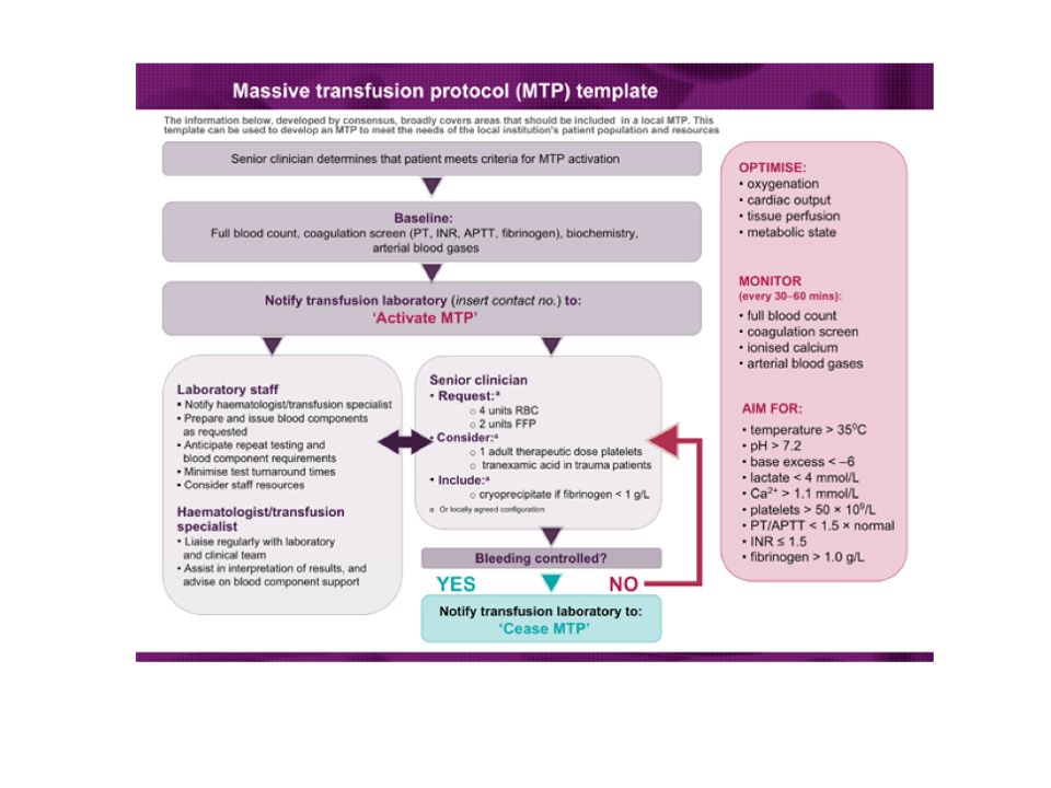
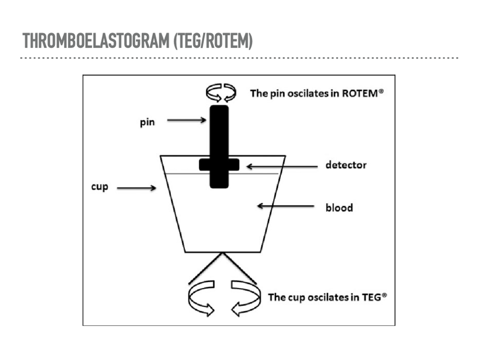
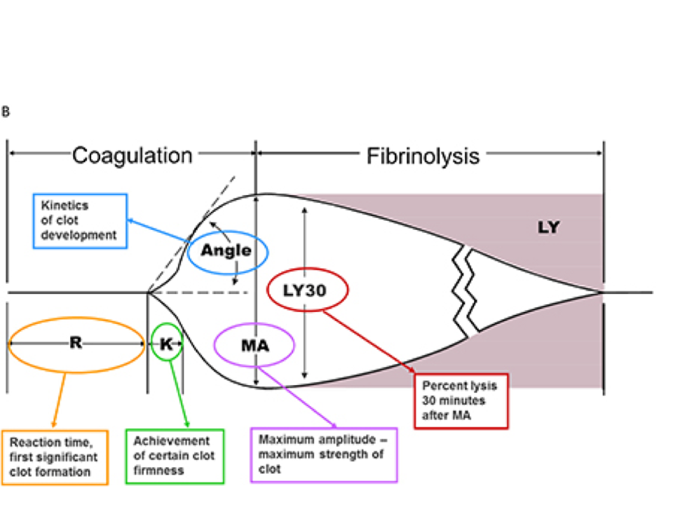
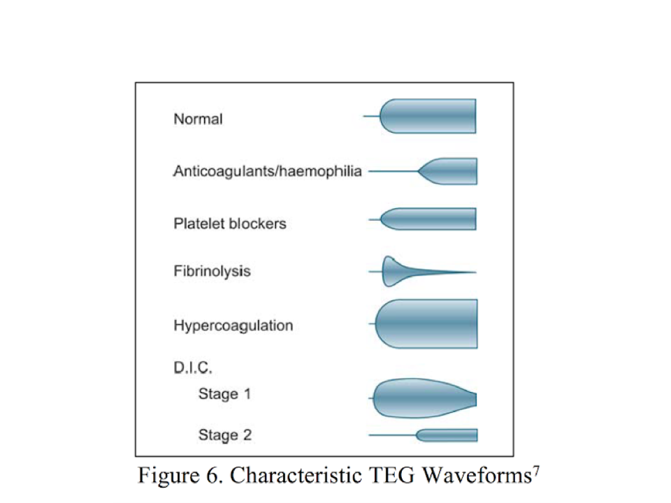
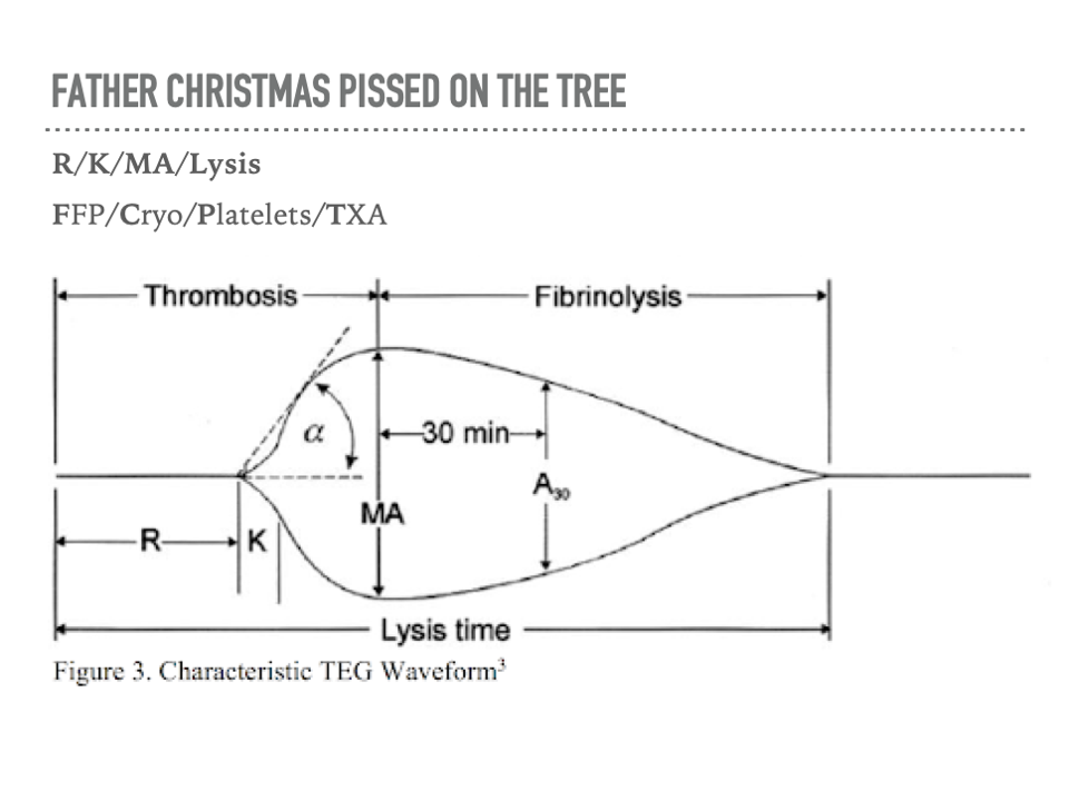
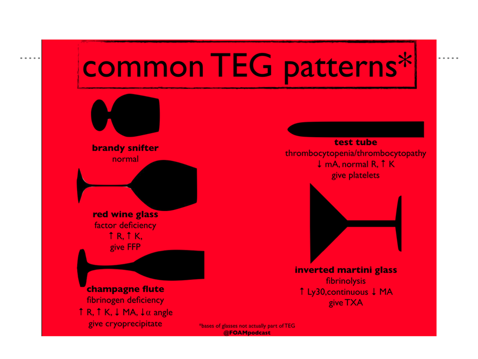
Joel’s talk summarised how to manage the shocked, bleeding trauma patient. This topic is expert level knowledge for ACEM trainees and comes up again and again in exams. The slides contain all of the key points so rather than me write them out here, have a flick through the slides below.

ECG of the Week
Thanks Sarah and Matt for this real case from recent weeks.
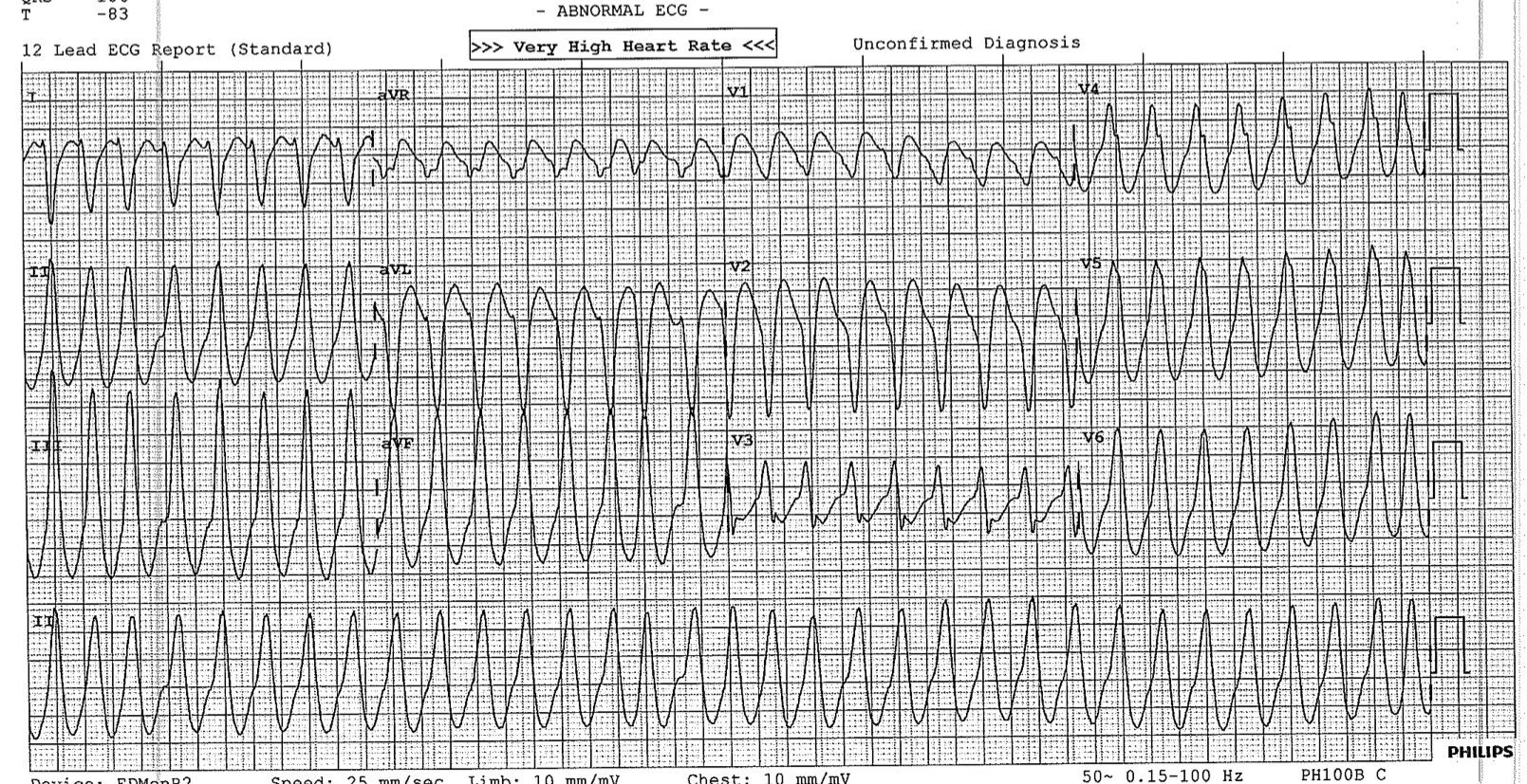
A 76 yo man was BIBA with the presenting complaint of dizziness and a systolic BP of 70mmHg. He had no chest pain.
Here is the initial ECG
ECG 1 - Arrival
Here we can see a regular, broad complex tachycardia. The differentials are VT, VT, VT and VT particularly in a 76 yo patient. The other differential is SVT with an abberrant conduction pathway but this is far less likely in this situation. There is a great list of how to differentiate between VT and SVT with an abberrant pathway on life in the fast lane.
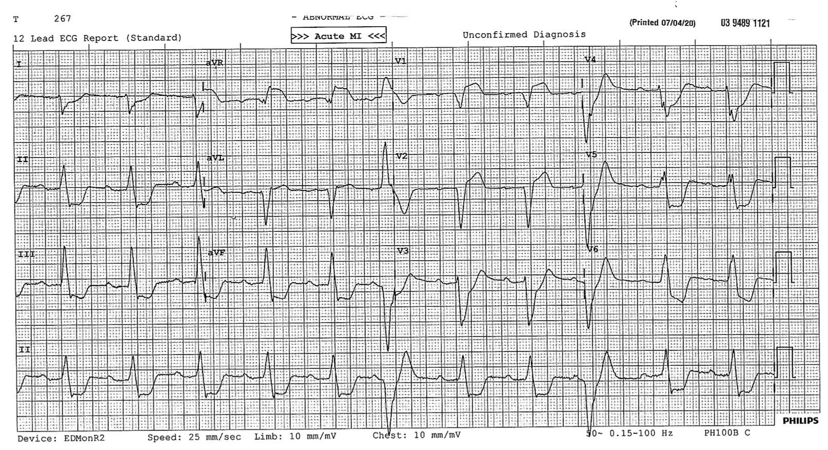
The patient was cardioverted with the next ECG as follows.
ECG 2 - Post cardio version
This ECG simply looks bad.
Abnormalities include:
1st Degree HB
LBBB
Broad QRS complexes >120ms
ST elevation aVR, V1, V2
Widespread ST depression
Several ventricular ectopic beats
In the setting of recent VT and haemodynamic instability this ECG screams out ischaemia and the cath lab was appropriately activated. Somewhat academic in this case, but a good teaching exercise, the modified Scarbossa Criteria can be used to help justify the decision for cath lab activation. The presence of any of the following suggests ischaemia and consideration of reperfusion therapy. Of note, these are still coming into acceptance with some cardiologists. They are present in European guidelines for cath lab activation but not yet in the US guidelines.
≥ 1 lead with ≥1 mm of concordant ST elevation
≥ 1 lead of V1-V3 with ≥ 1 mm of concordant ST depression
≥ 1 lead anywhere with ≥ 1 mm STE and proportionally excessive discordant STE, as defined by ≥ 25% of the depth of the preceding S-wave.
Out of interest, this patient turned out to have a normal angiogram, high troponins and is currently being investigated for myocarditis.
Thank you Sarah and Matt for the case.
If you have any good ECGs please forward them on to me.
Interesting VBG
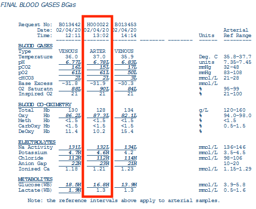
Following our blood gas discussion last week here is an interesting VBG to have a go at interpreting. Try the traditional method, try using strong ion difference, or try both. If we have time in the teaching session this week we can discuss.
Arbitrarily I have picked the middle gas and although labeled as arterial I suspect it is actually venous unless very hypoxaemic with pO2 of 61!
Perhaps the treating doctor can let me know?