
2 December 2020 - Paediatrics
Program
1000-1100: Morbidity and Mortality: Kirsten Cassidy
1100-1130: NAI - Ben S
1115-1130: Break
1130-1200: Oh no its a neonate!: Registrar talk - Jacqui M
1200- 1230: ECG/ image/ gas of the week - Maria, Mark and Jess, recap from 25/11: Mim
1230-1300: APLS quiz: Brendan
1300-1330: Inreach in the ED, geriatric registrar
1330-1400: Urogenital infections - Jess
1000-1200: OSCE Group
1315-1500: Simulation Session: Mim and Divya
ECG of the Week
No clinical context this week other than it was a real St Vincent’s patient.
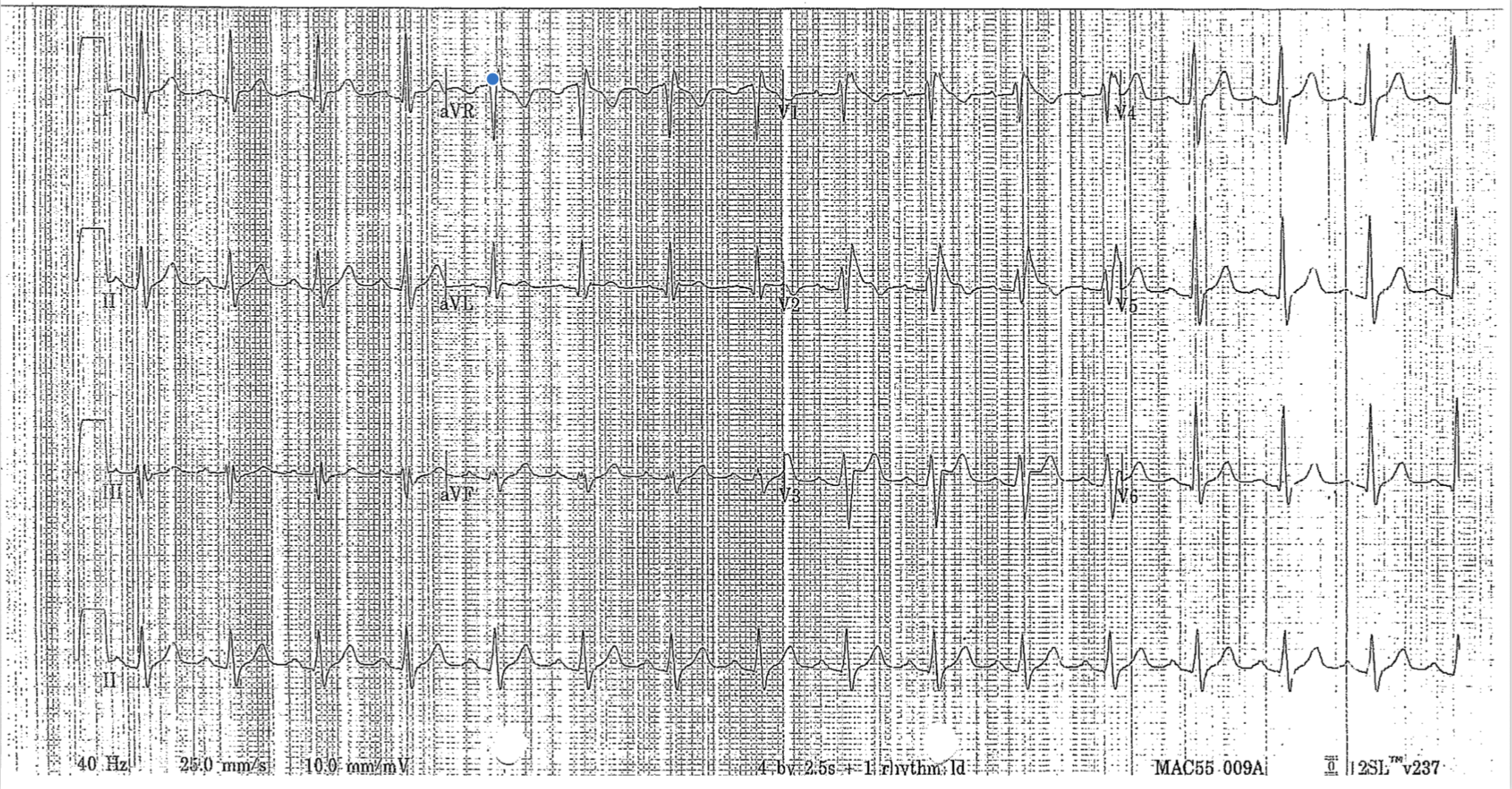
This ECG has the appearance of Type 1 Brugada Sign.
Remember that the ECG is not enough to diagnose Brugada Syndrome. To have Brugada syndrome you need to have a suggestive ECG and at least one clinical feature including:
Documented VF or polymorphic VT
Family history of sudden cardiac death at < 45 years of age
Coved-Type ECGs in family members
Inducibility of VT with programmed electrical stimulation
Syncope
Nocturnal atonal respiration.
For more details about Brugada ECGs and syndrome LITFL has a great review. https://litfl.com/brugada-syndrome-ecg-library/
Image of the Week
A six year old girl is brought into ED following a fall off a scooter onto her outstretched hands. She is complaining of pain in her right elbow.
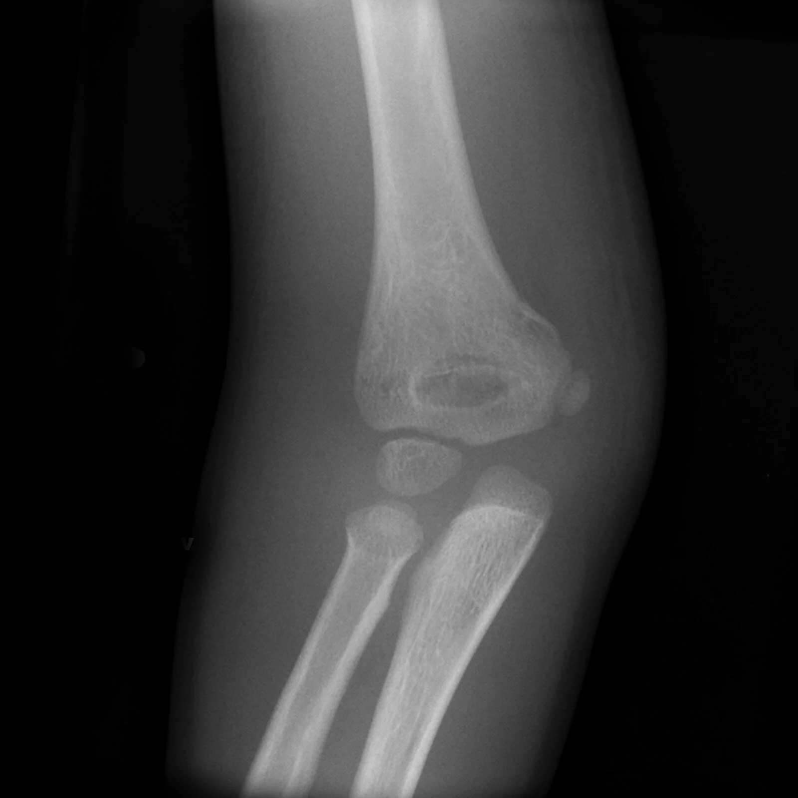
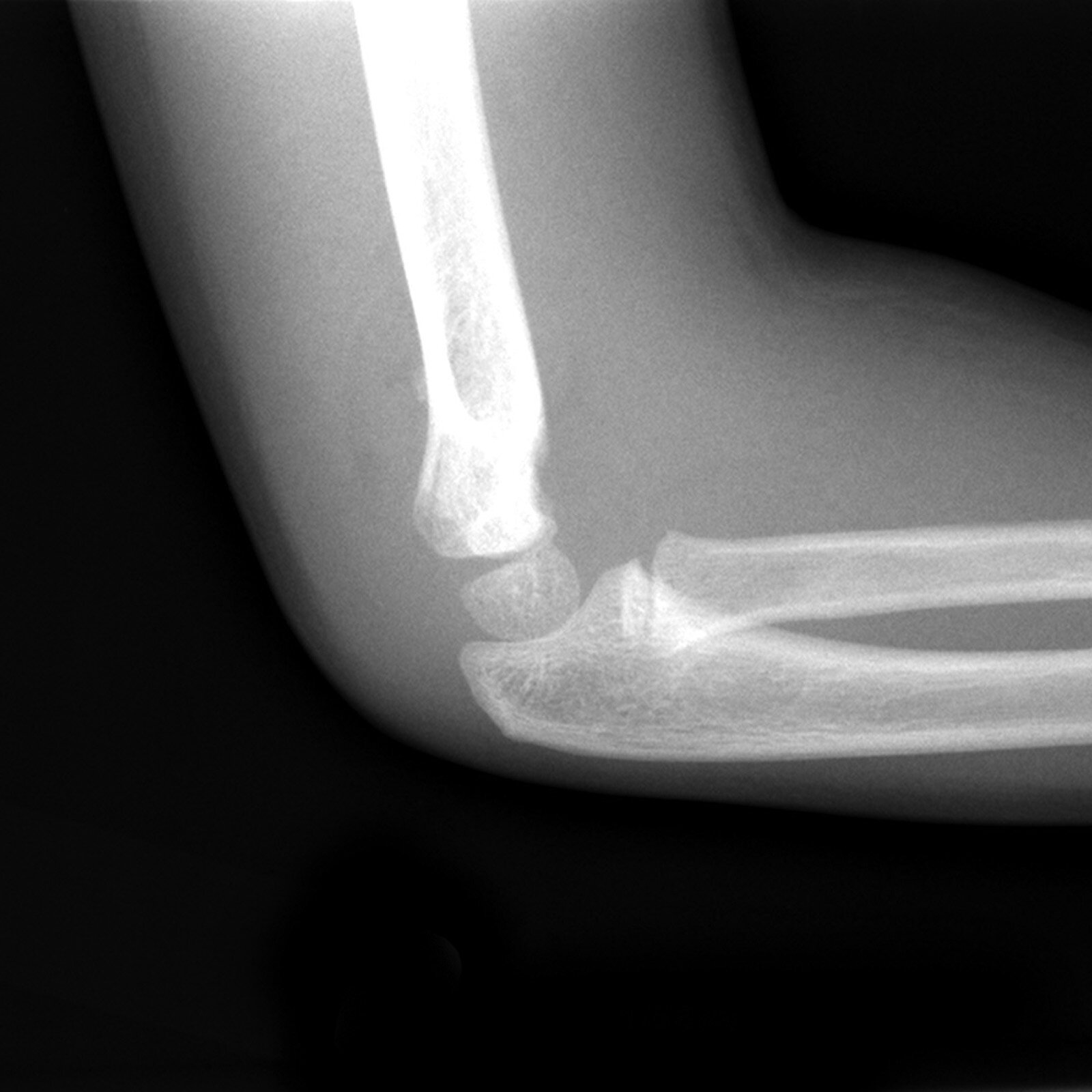
Image of the week this week shows a Gartland II supracondylar fracture. Remember to look for the anterior humeral line which should intersect with the middle third of the capitellum.
Other key features to look for on all paediatric elbow injuries include:
Anterior and posterior fat pads
Anterior humeral line and radiocapitellar line
Ossification centres (CRITOE pneumonic)
RCH has a great review of all common paediatrics fractures including supracondylar fractures which are the most common paediatric fracture about the elbow.
Gas of the Week
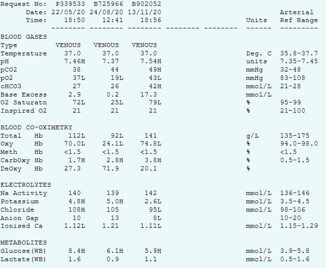
A 72 yo male was referred to the ED with a potassium of 2.7 mmol/L on outpatient bloods. He has no symptoms.
He has a history of metastatic SCLC with metastases to lung, nodes, liver and bone. His current treatment regime consists of carboplatin, etoposide and acetazolamide.
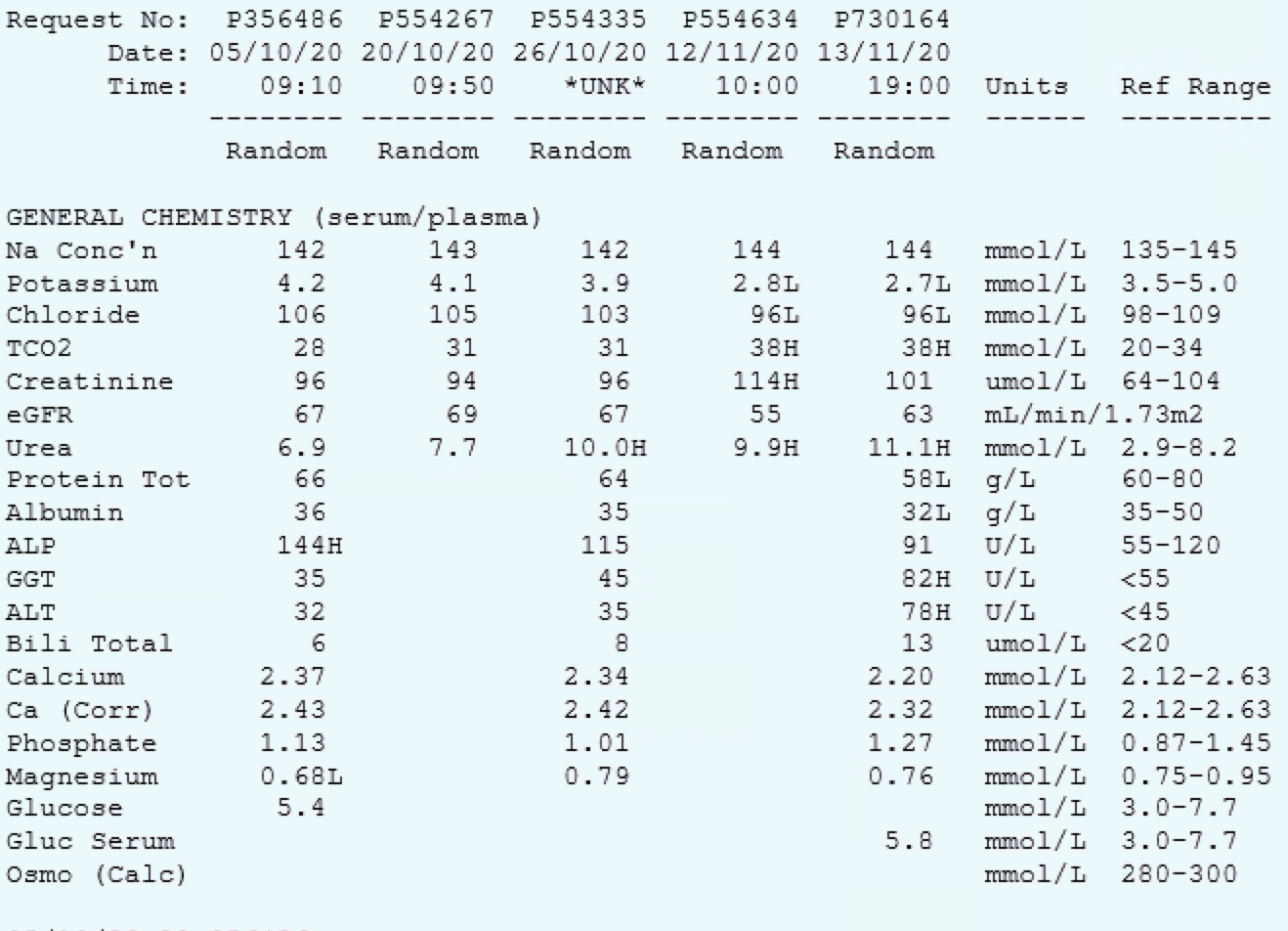
Key Findings
There is a metabolic alkalosis proses
Expected pCO2 = 0.7 x bicarb + 20 = 49
Hypokalaemia. Corrected K = 3.3
Fencl-Stewart
SID = 142+2.6 - 95 = 49. Therefore there is a hypochloraemic alkalosis with a chloride effect of +7
Lactate effect = 0
Albumin effect = +3
The measured base excess is +17 so there is another metabolic alkalosis contributing +7 to the base excess. This is likely to be a chloride unresponsive alkalosis. Causes of this include excess renin/aldosterone or extreme hypokalaemia.
Causes of metabolic alkalosis
G - GI acid loss - ? Vomiting secondary to cancer or chemotherapy, ? Diarrhoea secondary to chemotherapy
R - Renal acid loss such as diuretics, Bartter/Gitelman/ Milk-alkali syndrome - unlikely
O - Overdose of base - antacids, alcatives, HCO3 administration - no evidence of this
E - Endocrinopathy - ? No evidence of this
This patient ended up having an exotic ACTH secretion from their SCLS