
14 October 2020 - Miscellaneous
Program
1000-1100: Dermatological Emergencies: Aaron Robinson
1100-1130 - Recap from last week + ECG/ Gas/ Image of the week: Jess/ Mark/ Maria
1130-1145: coffee break
1145-1215: Common rashes in the ED: Tom
1215-1245 Envenomation update : Camilo
SMALL GROUP TEACHING:
1000-1200 - Fellowship OSCE Group
1300-1400: Primary preparation with Sarah
1300-1400: FW – last weeks tox and some others, Jac
1315-1445: Andy C and Tom: Airway Sim with Brendan
1300-1500: US with Mark Email Mark to secure your spot
Learning Points
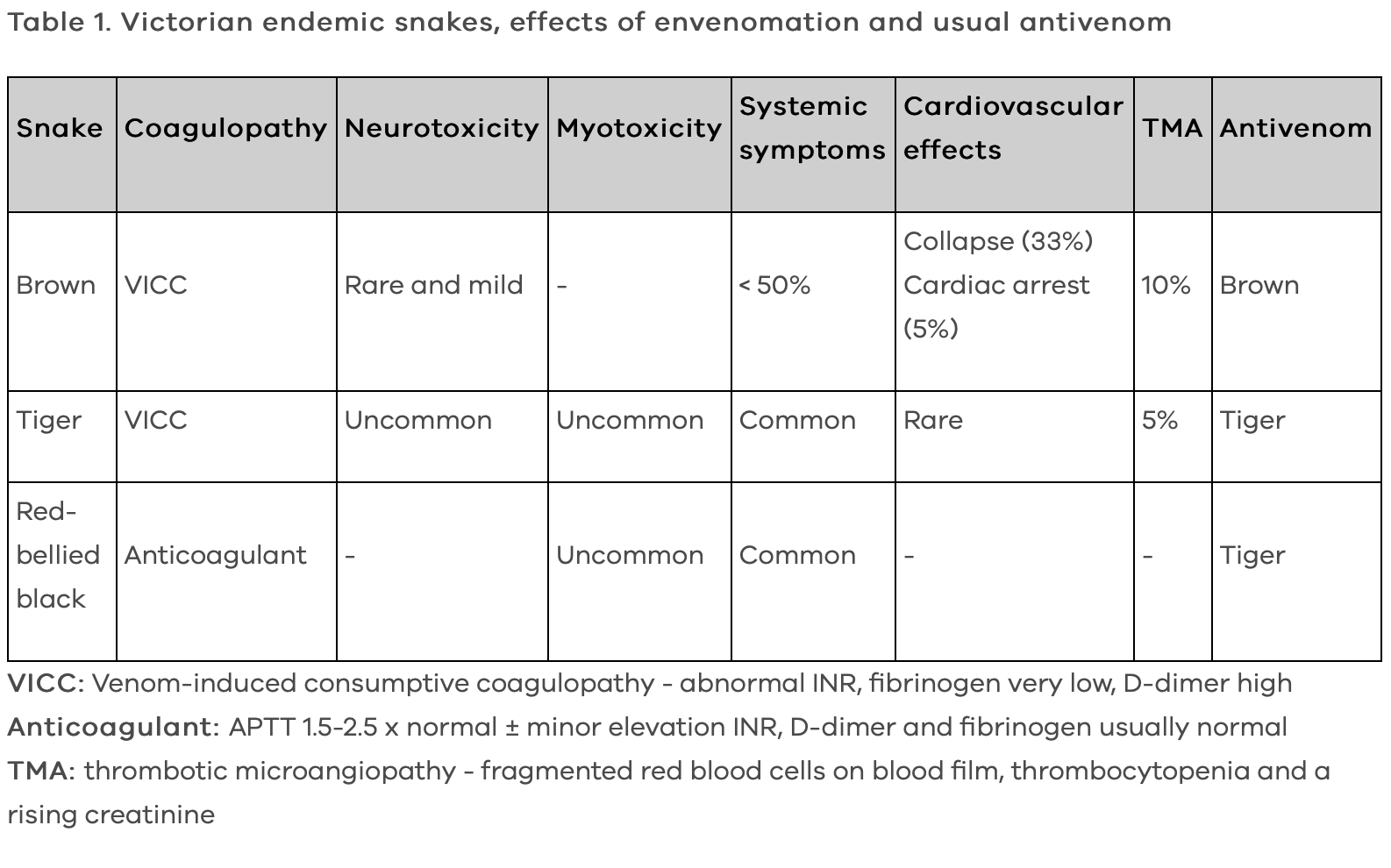
Snake Bites
This is a huge topic and is super important. Although we don’t see many snake bites at St Vincent’s knowledge of how to assess the patient with the suspected snake bite and management of envenomation is core knowledge for the emergency physician. It comes up on exams frequently.
Have a look at the Safer Care Victoria Page on snake bites. Here you can find management guidelines, patient information and up to date information.
Here are three key points to keep your patient’s safe:
Pressure bandages with immobilisation (PBI) save lives.
Discuss all snake bite patients with a toxicologist
Be prepared to manage anaphylaxis when giving anti-venom
ECG of the Week
Thanks Jess for taking over this segment and for providing this ECG
An 88 year old female presents from home with fevers and BP 102/74.
This is a really tricky ECG.
Let’s break it down into the objective findings first:
Irregularly irregular tachycardia, with rates up to 250bpm
Borderline right axis deviation
Variable QRS morphologies - some sinus RBBB QRS complexes, some ventricular ectopics, others
Variable atrial morphologies
Small QRS complexes in precordial leads (likely due to COPD diagnosis)
So what does this mean? In the context of a patient with COPD this is likely a picture of multifocal atrial tachycardia with ventricular ectopics. Differential diagnoses include atrial fibrillation (bu probably too fast), sinus tachycardia with atrial ectopy or atrial fibrillation with an aberrant pathway.
The treatment of multifocal atrial tachycardia is to treat the underlying diagnosis. It is a poor prognostic feature in patients with chronic disease.
Image of the Week
24F presents following a reported inversion injury of her ankle whilst intoxicated. An x-ray of her ankle and foot is shown below. Click on the images to enlarge
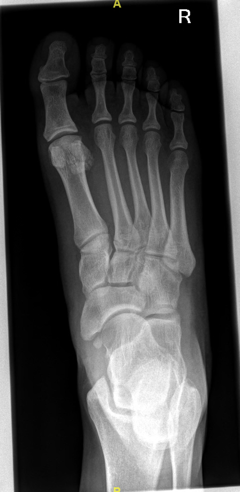
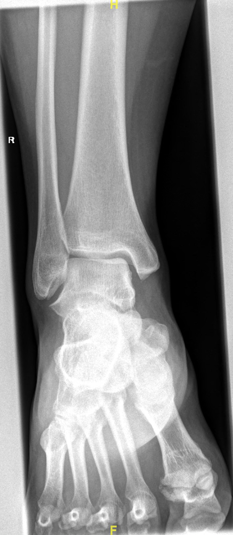
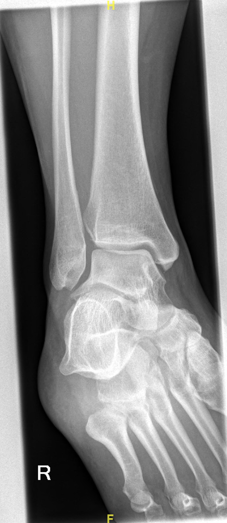
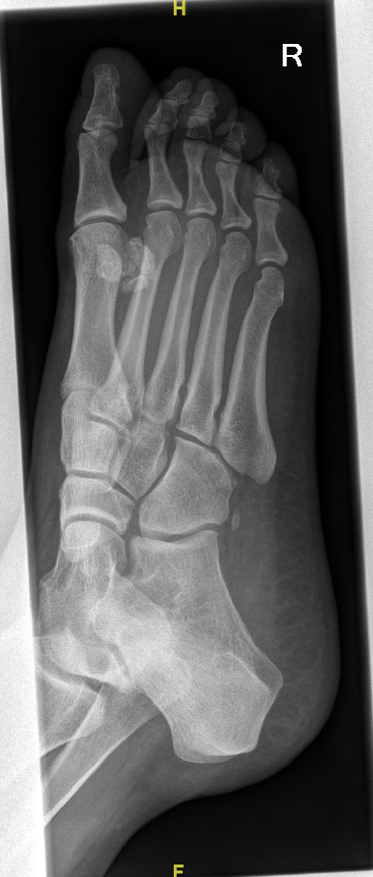
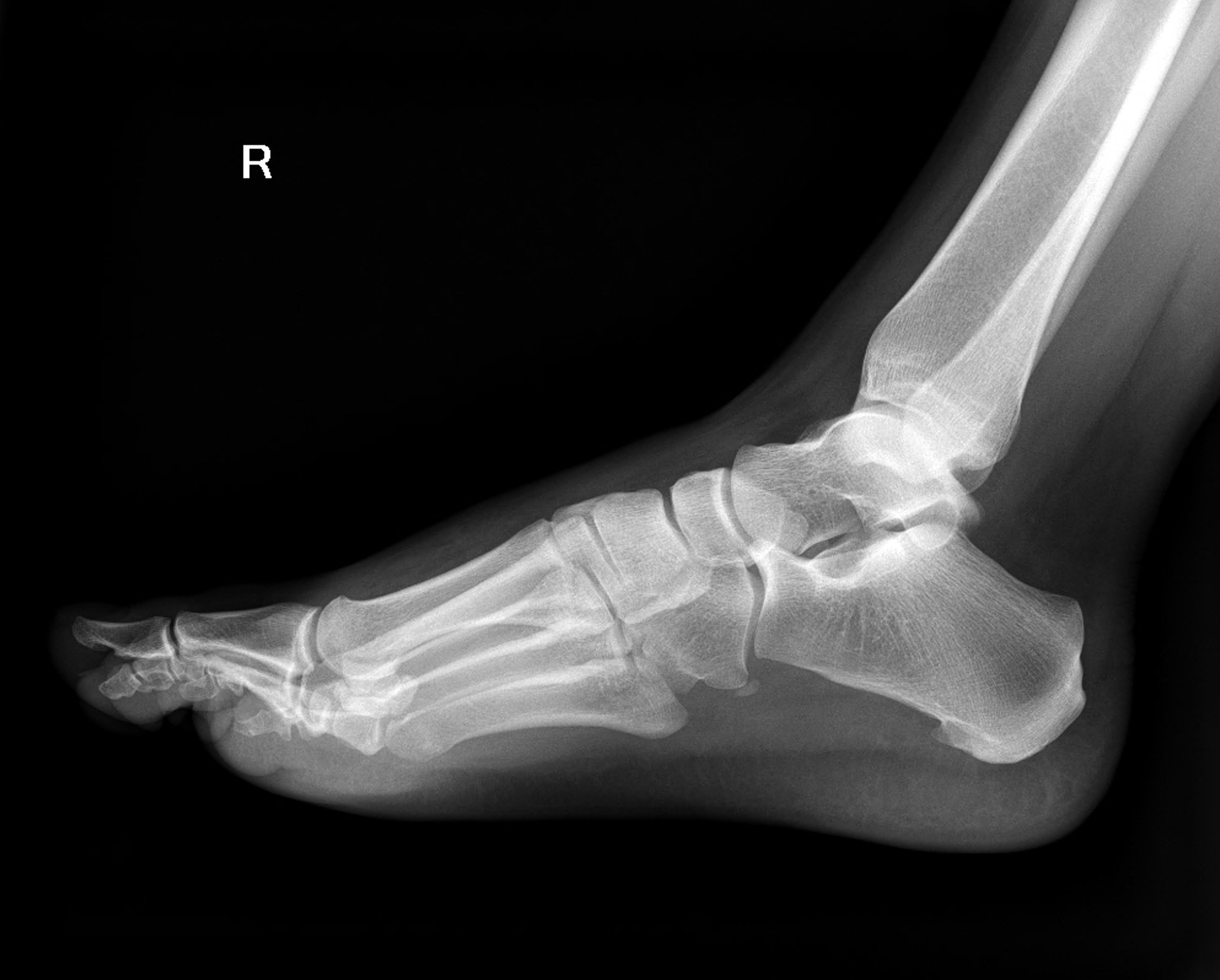
This image is concerning for a Lisfranc fracture-dislocation. The findings are subtle and further imaging is required.
The key finding is malalignment of the second tarsometatarsal joint, such as lateral displacement of the second metatarsal base as can be seen in these X-rays. There is a small fracture at the base of the second metatarsal commonly called the ‘fleck’ sign. Have a look at the radiopaedia link for further information. https://radiopaedia.org/articles/lisfranc-injury
The usual mechanism of injury is plantar flexion with external rotation.
Clinical findings
Inability to weight bear
Dorsal mid foot swelling
Plantar echhymoses
Gap between the toes
Inability to perform a single limb heel rise
When there is any suspicion of a lisfranc injury the patient requires a CT and possibly an MRI. Surgical management is sometimes required. Orthopaedic opinion is standard of care.
Gas of the Week
72 y.o. recent AVR, MVR, had been transferred back to rural hospital to recuperate, returns via ARV intubated, shocked.
Albumin 26.
Stops in ED ‘for a quick assessment’ prior to ICU.
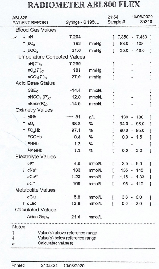
Primary acid-base problem: Low pH, low bicarb, base excess -15 so there is a moderate metabolic acidosis.
Fencl-Stewart Method
SID: 133 + 4 - 100 = 37. Therefore there is a hyperchloraemic acidosis contributing -5 to the base excess. ? renal impairment, ? Addison’s disease, ? steroid use
Albumin effect: Albumin should be 42 so it is down by 16. There is a mild hypoalbuminaemic alkalosis contributing +4 to the base excess
Lactate effect: -13
Sum effect is -14. This correlates to the measured base excess so there is no ketoacidosis or other unknown weak acid
Traditional method
AG = 133 - 100 - 12 = 21 (or 25 if using potassium). Therefore there is a HAGMA. The cause of which is most likely the raised lactate.
Delta ratio (21-12)/(24-12) = 9/12. There is therefore also a mild NAGMA (hyperchloraemia)
Winter’s Formula for expected pCO2 is 1.5 x 12 + 8 = 24. The actual pCO2 is 31 so there is a concurrent respiratory acidosis. In the context of a ventilated patient this effectively means ‘uner-ventilation’. We should increased the tidal volume by increasing the respiratory rate of this patient.
Further, there is hyperoxia. Do we need to decrease the FiO2? The current FiO2 is unknown so an Aa gradient cannot be calculated.
Putting it all together, this patient is shocked - ? sepsis, ? cariogenic. But you could probably guess that from the stem. Right?