
28 October 2020 - Geriatrics
Program
1000-1030: Recap from teaching 14/10/20 (Maria) and Alfred trauma 21/10/20 (Andy K), Hope’s case
1030-1100: Anaemia in the Elderly: Andy K (Postponed)
1100-1200: Indigenous Health Equity training
1200-1230: SVHED Trauma response - Emmeline
1230-1300: Break
1300-1330: Fractures in the elderly: Andy C
1330- 1400: ECG/image/ gas of the week - Jess, Maria, Mark
1000-1200 - Fellowship OSCE Group
ECG of the Week
84F presents with a labile BP. 199/105 to 82/60 and back up again to 172/86 with just 150mL normal saline.
No chest pain. Living in own home independently.
PMHx
IHD (nil PCI - medical management only)
DM II (OHGA)
OP + OA
NESB
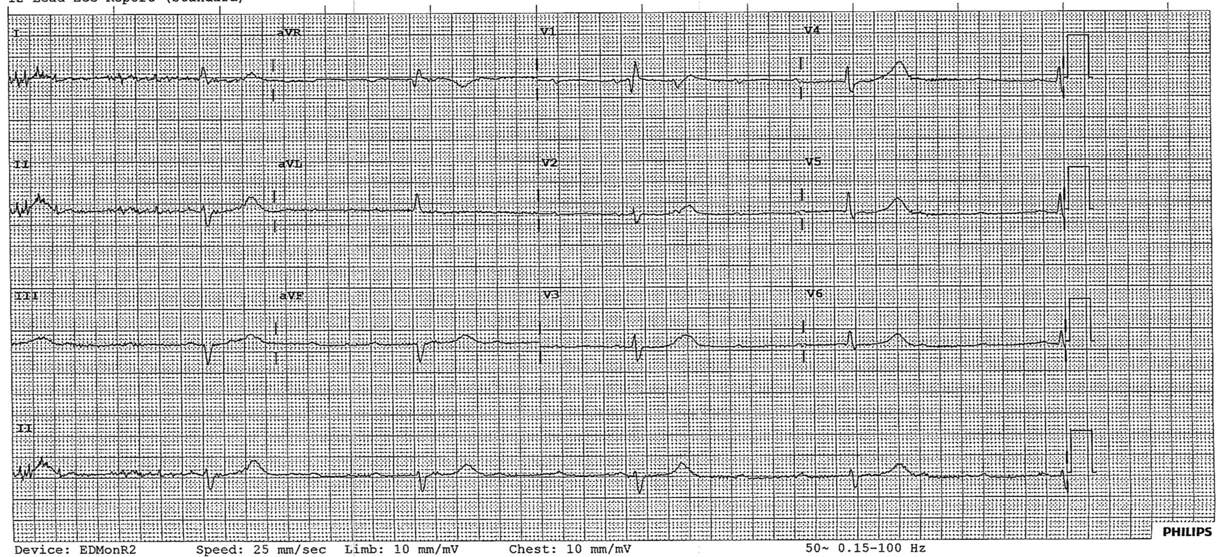
This ECG shows a complete heart block as there is no relationship between the p-waves and the QRS complexes.
There are regular p-waves at a rate of about 100bpm. The ventricular rate is approximately 36bpm
Image of the Week
A 47 year old female presents with acute epigastric and RUQ with vomiting. Her past medical history includes hypercholesterolaemia, psoriasis, psoriatic arthritis and depression. Her medications are rosuvastatin, methotrexate, folic acid, sulfasalazine and venlafaxine.
Click the images to enlarge
These images show pancreatitis. There is fat stranding and the borders of the pancreas are poorly defined. Further views would be needed to identify the presence of an obstructing gallstone. (Negative on ultrasound)
In this patient the patient’s medications were thought to be the cause. Remember the two most common causes of acute pancreatitis are alcohol and gallstones.
Gas of the Week
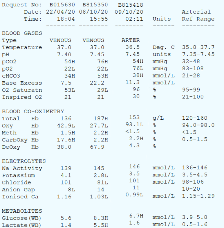
Have a look at the two later blood gasses.
The patient initially presented on 22/04 with ‘collapse’ for investigation. They had a neuro workup and was discharged without a diagnosis.
The patient then presented on 8/10 in status epilepticus
PMHx: known seizure disorder, chronic pain, PTSD, polypharmacy
The paramedic handover given was ? 2 hours of seizure - complex partial on background of known epilepsy. There was no evidence of trauma. Noted to be febrile with AV
The patient was treated with 15mg IM midaz and 10mg IV Midas with AV and no improvement in seizure
On arrival -
A - own, drooling
B - RR 40, Sao2 99% 2L
C - HR 82, BP 120/50
Febrile 38.8 - primary or secondary ?
GCS 3
Pupils 4mm not reacting, deviated to R
R sided upper limb, lower limb and abdo tonic clonic seizure
The VBG at 15:15 was on arrival. The ABG at 02:11 was taken hours after intubation.
Albumin = 38
Interpretation
Venous gas
Overall normal pH but nearly an alkalaemia.
On first glance there is a metabolic alkalosis and a respiratory acidosis
Expected pCO2 = 0.7 x 53 + 20 = 57
Although this is a venous gas there is likely a concurrent respiratory acidosis
Causes of metabolic alkalosis - ? Vomiting although not in the history. ? Renal acid loss or endocrine cause
Causes of respiratory acidosis - hypoventilation, ? Secondary to seizure.
There is also a high lactate that could be secondary to the seizure or hypoxia.
There is also severe hypokalaemia requiring urgent replacement
Fencl-Stewart
SID = 145+3-81 = 67 so there is a hypochloraemic metabolic alkalosis contributing +25 to the base excess
Albumin effect = +1
Lactate effect = -5
The sum effect is 21 which matches the measured base excess.
Arterial gas
Overall normal pH
Moderate metabolic alkalosis
Expected pCO2 = 0.7 x 38 + 20 = 46.6 so there is still a respiratory acidosis present
Fencl-Stewart
SID = 146+4 - 101 = 49. There is a hypochloraemic metabolic alkalosis contributing + 7 to the base excess
Albumin effect = +1
Lactate effect = -1
Sum effect is +7 so there is another +4 unknown metabolic alkalosis
Aa gradient = roughly 70 so there is a raised Aa gradient suggesting VQ mismatch. ? Aspiration ? Atelectasis ? Pneumonia
Potassium has been improved from previous gas. Lactate improved. Acid base status improving.