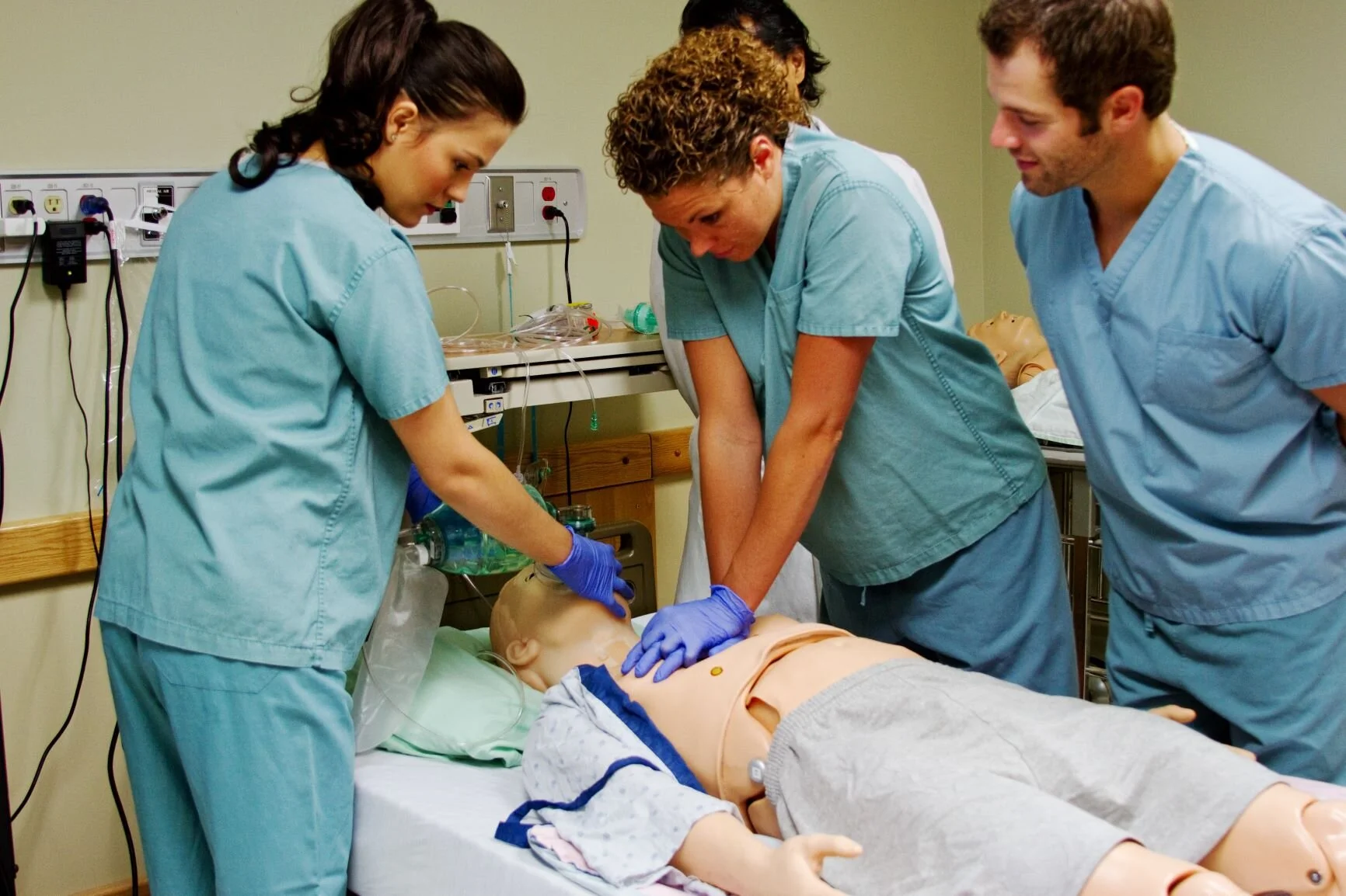
4 August 2021 - Advanced Life Support
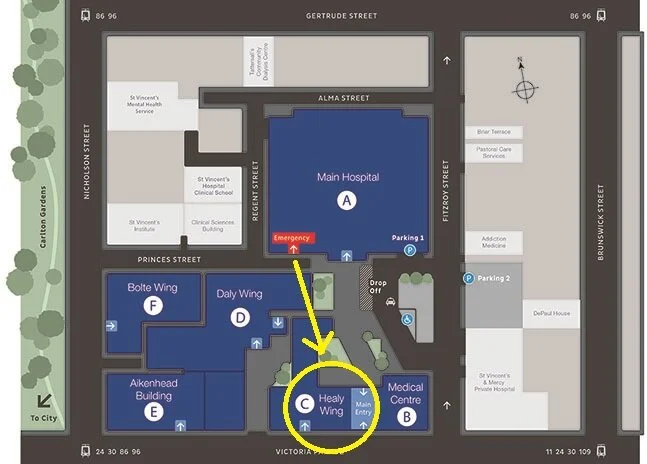
Teaching this week will be in the Teaching Lab in the Education Centre on Level 3 of Building C (Healy Wing).
Program
1000—1015: Welcome and introductions
1015—1030: Kahoot Recap — Gemma Ridley
1030—1045: ECG and Image of the Week — Ben Lee
1045—1100: Research and Quality Portfolio Allocation — Hamed Akhlaghi
1100—1115: EDNAV Update — Jacqueline Maplesden
1115—1140: Emergency Treatment of Hyperkalaemia — Rex Gavan
1140—1215: Lunch and coffee break (no coffees in the lab sorry)
1215—1315: Skills sessions (30 minute rotations) — split into 2 groups
ALS scenarios
Procedural practice — CVC insertion, IO insertion, cricothyroidotomy
1315—1430: Further procedure practice for junior registrars 1:1 and Orientation —Jacqueline Maplesden and Sarah Ward
1315—1430: Fellowship Written Group — Alex Handrinos
1315—1430: Fellowship OSCE Group — Brendan Morrissey
ECG of the Week
60M presents with slight chest pain, slightly SOB, telling you he ‘feels fine’. BP 140/80.
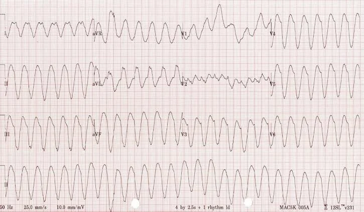
1) Describe and interpret the ECG
Broad complex monophasic tachycardia -- suggestive of VT
2) Name 5 conditions which can predispose an individual to this pattern
Most commonly IHD! However when listing conditions, think "cold, cold heart" :
Channelopathy (e.g. Brugada)
Other (electrolytes)
Long QT
Drugs (e.g. cocaine)
Cold = Hypothermia
Heart = Structural (e.g. IHD, HOCM, arrhythmogenic right ventricular cardiomyopathy)
When it comes to drugs that cause a long QT, use this acronym to be an “ACEM IDOL” :
Amitriptyline and other TCAs
Clarithromycin and other macrolides
Electrolyte imbalances - low K, low Mg, low Ca
Methadone and metoclopramide
IDOL = droperIDOL, haloperIDOL
Another way to remember drugs that cause long QT is by class of drug:
Antidepressant — e.g. amitriptyline
Antibiotics — i.e. macrolides like azithromycin
Antipsychotics — e.g. droperidol, haloperidol
Anti-emetics — e.g. ondansetron
Anti-arrhythmics — e.g. procainamide, amiodarone, sotalol
3) Name 3 cardiac mechanisms that can bring about VT
Re-entry (most common)
Triggered activity -- early or late after-depolarisations (e.g. torsades de pointes)
Abnormal automaticity -- abnormal impulse generation by a region of ventricular cells
4) How would you manage this patient?
IV amiodarone 5mg/kg (max 300mg) or IV sotalol 80mg
Cardiovert if pharmacotherapy fails or becomes haemodynamically unstable
Pacing if cardioversion fails
Identify and treat cause, e.g. Mg for long QT
Image of the Week
You are attempting direct intubation. Upon inserting your MAC blade into the vallecula you see this:
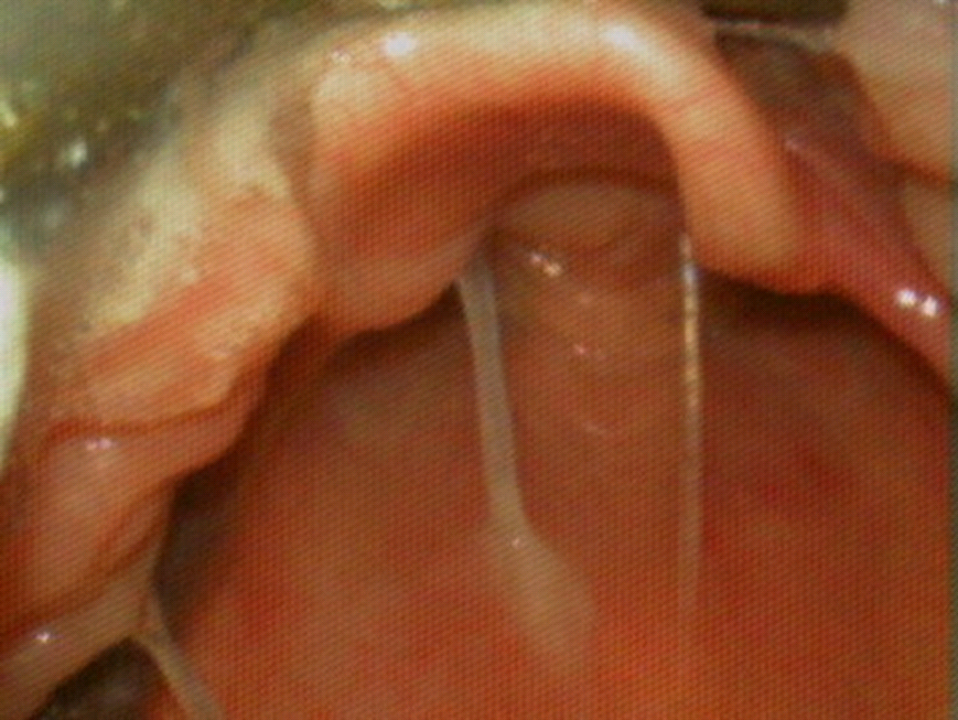
1) What intubation view category are you seeing here? Explain your answer by describing the structures you can see.
Modified Cormack and Lehane Grade IIb (arytenoids only just visible)
2) What other intubation views are possible? What structures can be seen in each?
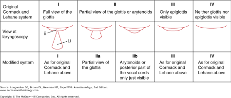
Grade I: Full view of glottis
Grade IIa: Partial view of glottis
Grade III: Only epiglottis visible
Grade IV: Neither glottis nor epiglottis visible
3) Without removing your MAC blade or inserting any adjuncts, what positional changes could you make during this intubation attempt to improve your view? Name 3.
Increase pulling laryngoscope towards the opposite corner of the ceiling
Backward, upward, rightward pressure (BURP)
Hyperflex neck
Lift head off the bed with your right hand, then keep it there with the strength of your left hand holding the laryngoscope or by bracing the patient's head against your abdomen
4) If the above are unsuccessful, and you are unable to intubate, what changes could you make in your 'Plan B' intubation to have success?
Use a bougie
Use a videolaryngoscope
Use a hyperangulated blade
Refer to a senior operator or anaesthetics