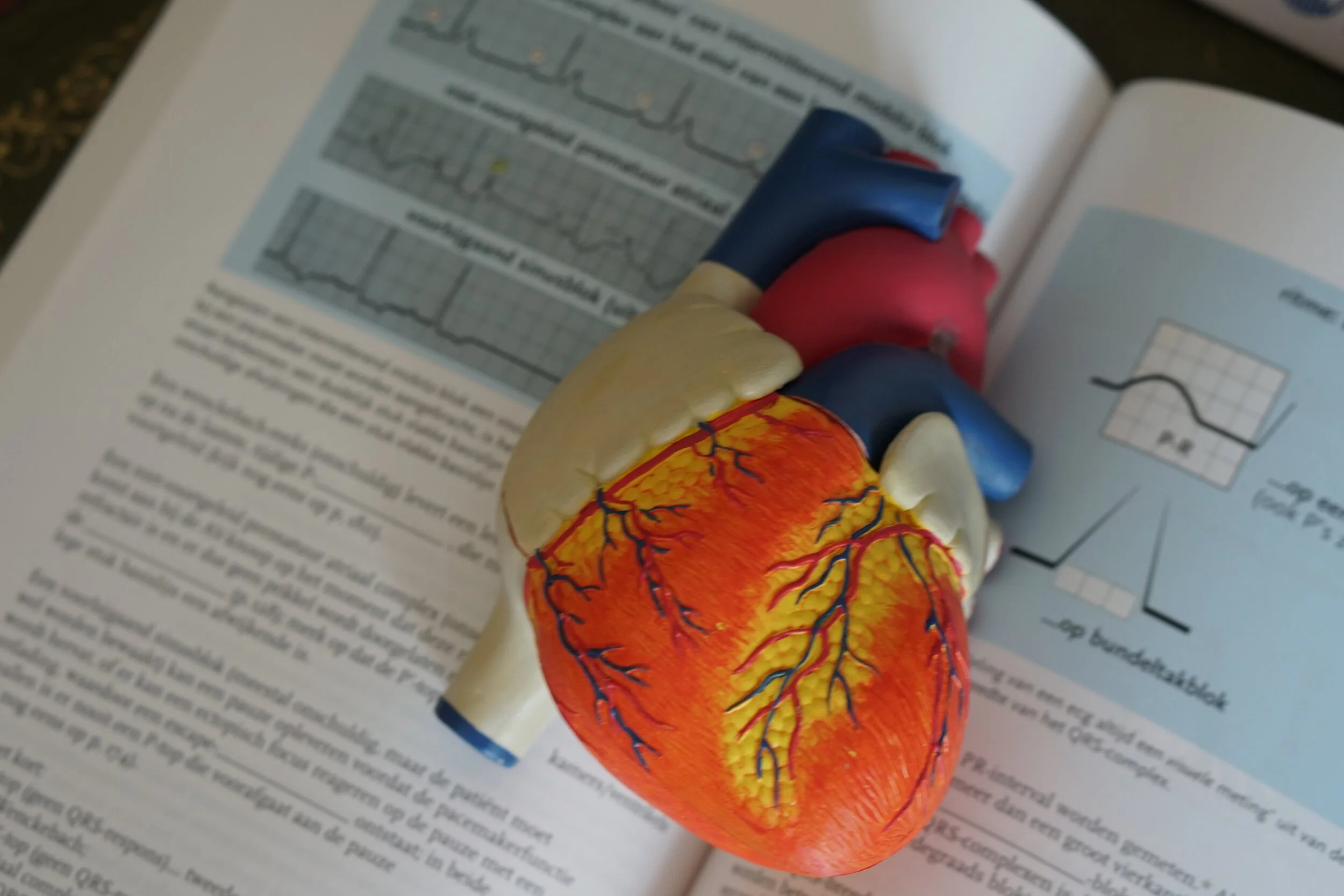
3 March 2021 - Cardiovascular II
Program
1000-1005 – Welcome
1005-1030 – Case Presentation – Ben Sinnett
1030-1130 – STEMI and ACS – A/Prof Andrew Macisaac (SVHM Director of Cardiology)
1130-1140 – Break (NB: unfortunately no coffees allowed in the Teaching Lab)
1140-1220 – Journal Club – Ben Lee and Rex Gavan
1220-1225 – Image of the Week – Ben Lee
1225-1230 - ECG of the week – Ben Sinnett
1230-1300 – Lunch
1300-1430 – Cardiology small group practical sessions (rotating through as per last week)
Cardiac simulation
Pacing and Defibrillation
POCUS Echo
ABGs
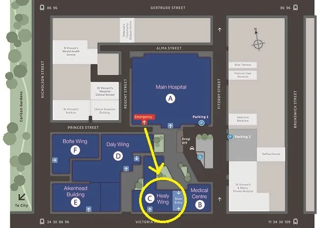
Teaching this week will be held in the Teaching Lab in the Education Centre on Level 3 of Building C (Healy Wing).
Key Learning Points
Thanks to Sarah Simons for this week’s teaching pearls.
Acute coronary syndrome (Medical Journal of Australia summary 2016):
https://www.mja.com.au/system/files/issues/205_03/10.5694mja16.00368.pdf
Negative troponin is a powerful prognostic indicator of a good outcome
Best practice is ECG obtained within 10 minutes of first patient contact and primary PCI within 90 minutes
Aim for SpO2 >93% in patients presenting with ACS (oxygen is a drug in itself)
Pacing
When pacing for unstable bradyarrhythmia, ensure cardiac monitoring with three lead ECG on a separate monitor from the pacing leads
Hypothermia as a contraindication for pacing
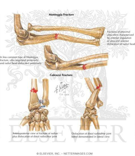
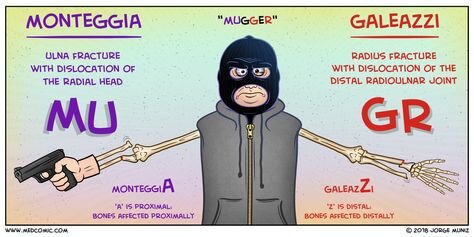
Monteggia vs Galaezzi fracture - what’s the difference?
Differentiating types of data
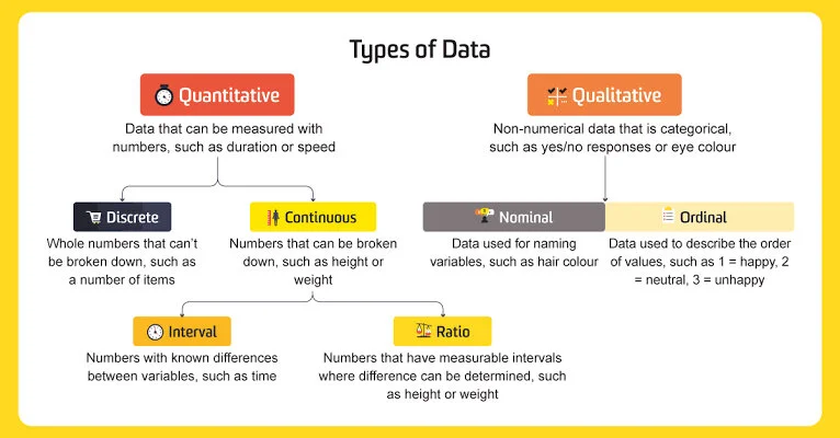
Image of the Week
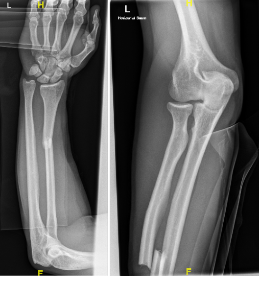
Describe the abnormalities.
Galeazzi fracture = Distal 1/2 or 1/3 radius fracture with distal radio-ulnar joint dislocation
This type of injury is often confused with another eponymous name. What is it, and can you describe this second injury?
Monteggia fracture = Proximal 1/3 ulnar fracture with radial head dislocation
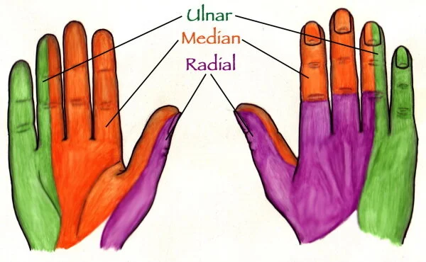
If a patient attended with this injury, how would you assess nerve function in the hand?
Ulnar nerve: Sensation over 1.5 fingers on the ulnar side of hand (volar and dorsal)
Median nerve: Sensation over palm, inner aspect of thumb, and volar aspect of first 3.5 fingers
Radial nerve: Sensation over back of hand
Checking power: Ask patient to make the “OK” sign
ECG of the Week
An 83yo lady presents with fatigue and BP 128/69 at triage. She has a past medical history of paroxysmal AF (secondary to an episode of pericarditis), COPD, HTN and RA. She takes diltiazem and flecainide.
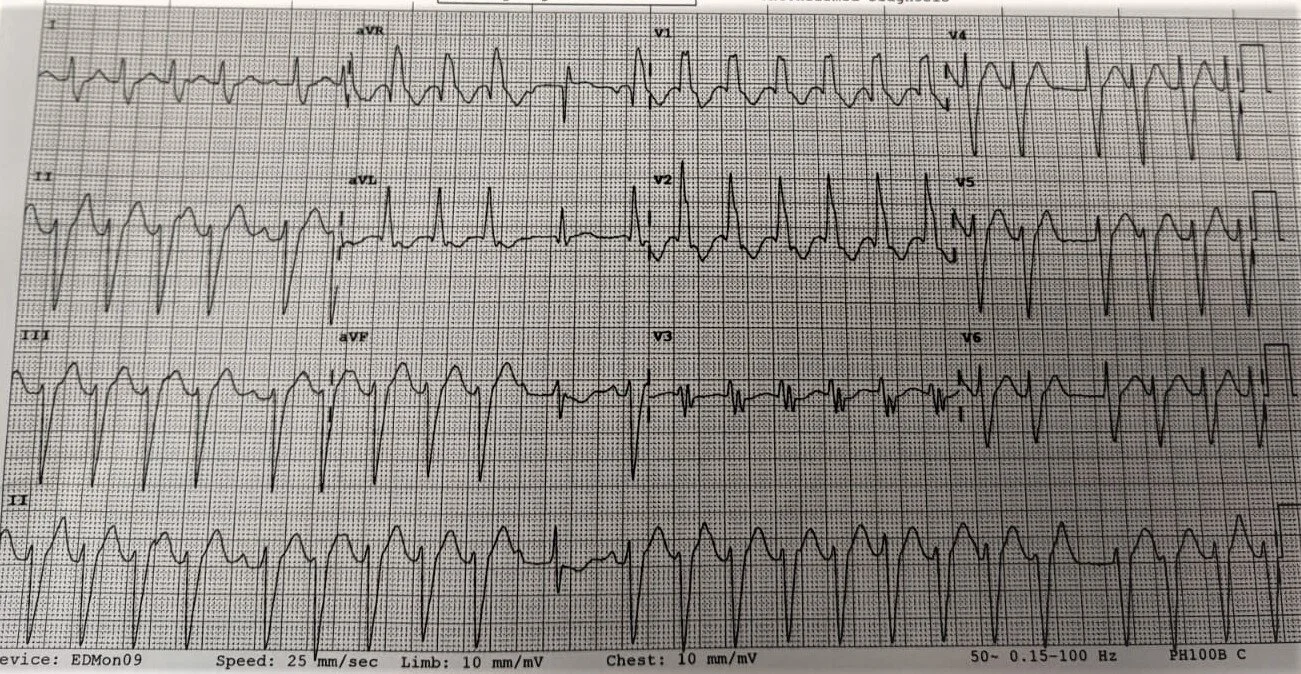
Describe the ECG.
Wide complex tachycardia at 140-150bpm
Irregular rhythm
Left axis deviation with RBBB (i.e. bifascicular – LAFB + RBBB)
P waves are intermittently present (see 5th beat in limb leads)
Prolonged QT interval
Anterior T wave inversion (V1 and V2)
10th beat looks like a capture beat
No delta waves
Is the rhythm of atrial or ventricular origin? Which aspects of the ECG support this?
80% of wide complex tachycardias are VT. This increases to >90% if the patient is >50yo and/or has a history of ischaemic heart disease.
To stratify any arrhythmia, begin with three questions:
Is the rhythm regular or irregular?
Is it fast or slow?
Are the QRS complexes broad or narrow?
In this case, the rhythm is irregular, fast and broad. Differentials include:
AF with a bundle branch block (aka "AF with aberrancy")
AF with WPW (accessory pathway)
Atrial flutter with variable AV conduction + aberrancy
Potassium or toxicological causes, e.g. TCA overdose
Polymorphic VT / Non-sustained VT
However if the rhythm was regular, fast and broad, differentials would be:
Sinus tachycardia with aberrancy
SVT with aberrancy
Accelerated idioventricular rhythm
Monomorphic VT (VT is usually caused by a stable re-entrant pathway, so has a consistent R-R interval.)
Diagnosis: This patient was treated as having atrial flutter with RBBB by Cardiology
Clues to final diagnosis:
Consistent amplitude counts against polymorphic VT
P waves = not seen in AF. Intermittent presence of P waves is suggestive of atrial flutter with variable block
Wide complexes conferred by a co-existing RBBB
Assuming a stable blood pressure, how would you attempt to correct the rhythm? How would this change if the patient was unstable?
The risk is in missing an accessory pathway (e.g. AF/flutter + WPW) because giving an AV nodal blocking drug like adenosine, beta blockers and calcium channel blockers can precipitate ventricular dysrhythmias and cardiac arrest. Ideally you’d check to see if the patient had an older ECG to compare with to make sure they don’t have WPW.
If stable:
- If 100% sure of the atrial flutter diagnosis, treat as you otherwise would. Treat cause if possible, rate control (in this case, the patient is on flecainide and diltiazem), optimise K 4.5-5.0 and Mg >1.0, seek Cardiology input.
- If unsure, treat as VT. Give amiodarone 5mg/kg and DCR.
If unstable: DCR