10 March 2021 - Respiratory
Program
1000-1030 — The Physiology of Common Respiratory Complaints - Laura Burns
1030-1045 — Leadership Needs Analysis and Skills workshop - Isobel Ursich, SVHM Learning & Development Coordinator
1045-1055 – Recap of last week’s teaching - Rex Gavan
1055-1100 — Break
1100-1130 — Clinical Decision Rules in the Work-up of PE - Jonathan Leslie
1130-1140 — ECG of the Week - Jannet Chan
1140-1150 — Image of the Week - Ben Lee
1150-1200 — Break
1200-1300 — Palliative Care - Tom Whelan, Palliative Care Nurse Practitioner, SVHM Centre for Palliative Care
The communication skills teaching resources mentioned in the Palliative Care talk can be found here (search: ‘palliative’). The site requires you to sign up, but it’s an extremely quick process, and there are lots of really helpful teaching resources covering a cavalcade of Gen Med topics.
1300-1400 — Team Leadership OSCE Session 1 - Brendan Morrissey
1400-1415 – Registrar Forum - Mim Scharkie and Phil Ward
Potentially also a joint session with ICU!
Teaching this week will be held on Zoom.
https://zoom.us/j/5981463478?pwd=RThaZzNkL2w5bldBMlNHeE1zZFFaZz09
Meeting ID: 598 146 3478
Passcode: 2021
Image of the Week
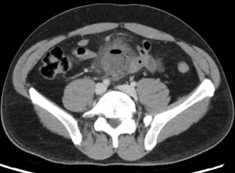
Describe this image and the abnormalities seen. Try to identify all structures in the scan (you don’t have to list them).
AP view of an abdominal CT with contrast
In the centre of the pelvis is a ring enhancing lesion with profound fat stranding
Gas locules seen within this structure and one small extra-luminal air pocket
What vertebrae are we seeing in this image? Explain your choice.
L5
Below bifurcation of aorta (L4)
Not a part of the sacrum
What are your differentials for the appearance of this CT scan?
Appendiceal abscess
Post-op abscess
Meckel’s diverticulitis
Diverticulitis
IBD abscess
Neoplastic lesion
ECG of the Week
49 yo male presented to ED with chest pain for 2 hours.
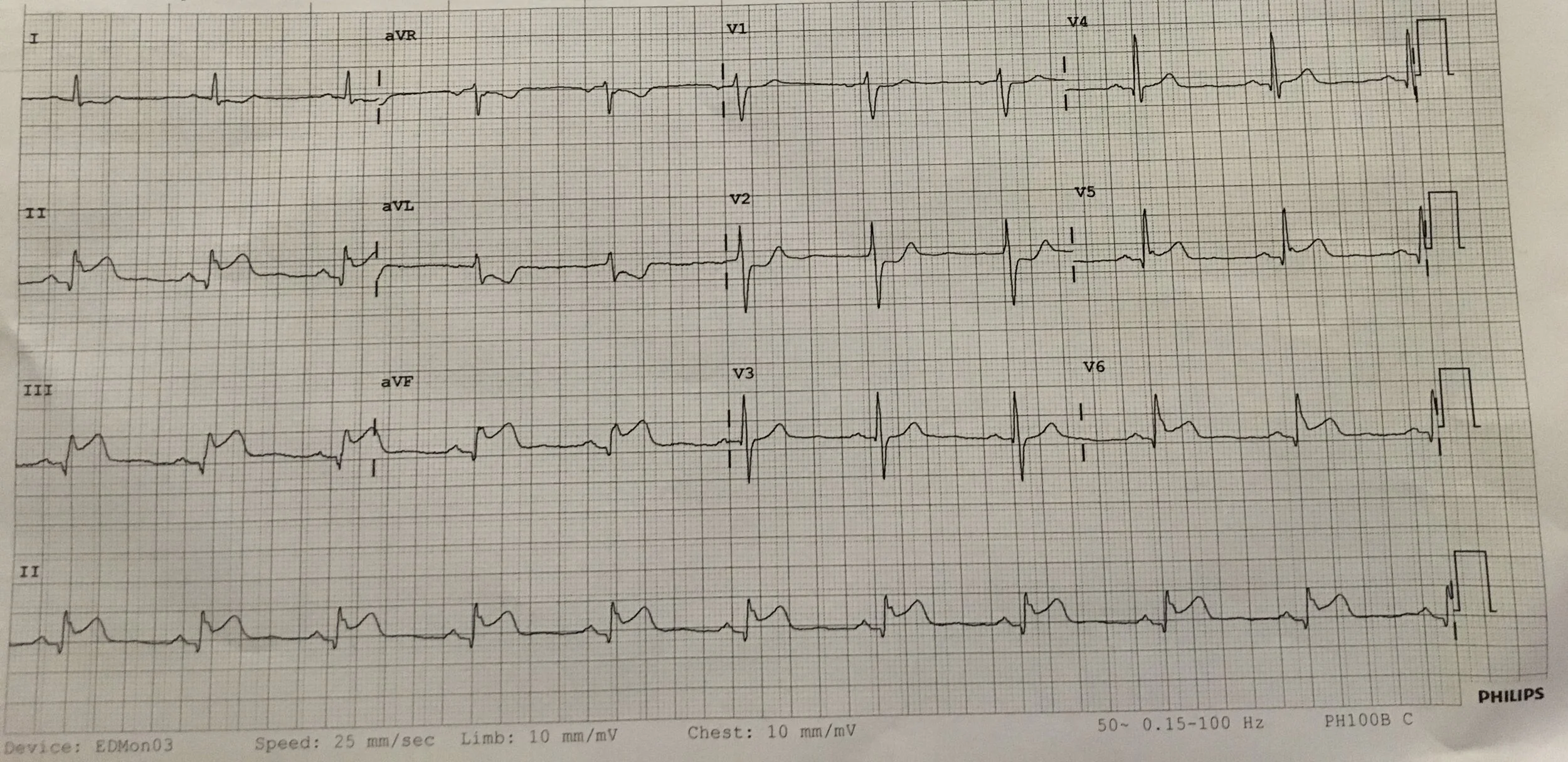
Describe 3 abnormalities.
ST elevation in inferior leads = Inferior STEMI
ST elevation in III more than II = Suspicious for right ventricular STEMI
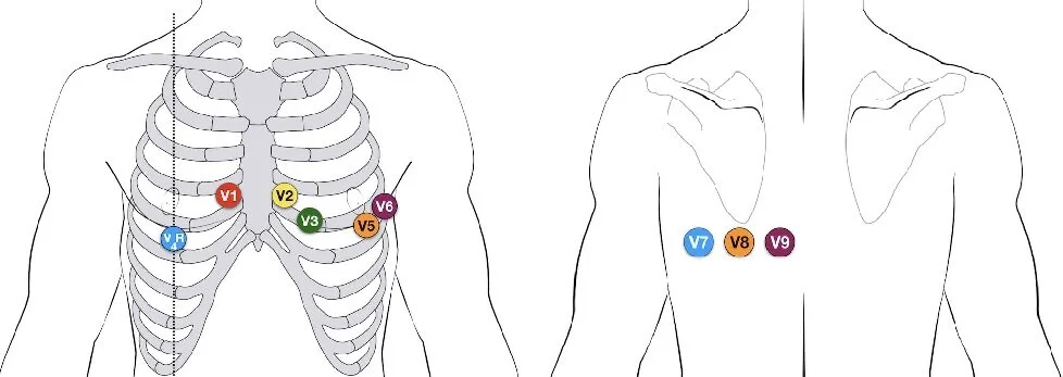
ST depression mainly in V2 = Suspicious for posterior STEMI
Confirm right-sided and posterior STEMIs with right ventricular ECG (below on left) and posterior ECG (below on right):
List 4 management priorities.
Code STEMI
PO aspirin 300mg, PO ticagrelor 180mg, IV heparin 5000 units
IV morphine 2.5-5mg for analgesia, provided SBP >100mmHg
Given right ventricular STEMI, aim SBP >100mmHg or MAP >60 with judicious fluid bolus 10-20mL/kg and/or inotropes
Name a medication you would avoid giving.
GTN — right ventricular STEMI relies on preload to maintain stroke volume