
17 March 2021 - Acute Behavioural Disturbance
Program
CONFERENCE ROOM
1000-1130 — Code Grey Training - Tiff Plummer and Security
1130-1140 — ECG of the Week - Daniel Coghill
1140-1145 — Image of the Week - Sarah Simons
1145-1215 — Lunch
1215-1245 — Organic vs Non-organic Psychosis - Ezra Limm
1245-1255 — Recap of Last Week’s Teaching - Sarah Bannerman
MOVE TO TEACHING LAB
1300-1330 — Drugs in Acute Behavioural Emergencies - Daniel Coghill
1330-1500 — Acute Behavioural Emergency Simulation
You may like to read up on the management of Acute Behavioural Emergencies prior to the session:
Better Safer Care Victoria’s Clinical Guidance for Acute Behavioural Disturbance
NUEM Blog’s Emergency Medicine Resident Education on Verbal De-escalation Strategies
All the best to our colleagues sitting the FACEM OSCEs this week!
Teaching this week will begin in the Conference Room in the Education Centre on Level 3 of Building C (Healy Wing).
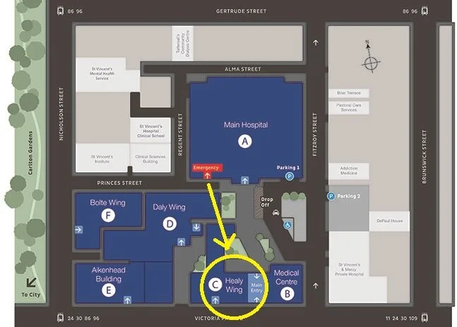
Image of the Week
79yo lady presents to your ED within 6 hours after a syncopal episode towards the end of a 4km walk. No head strike. She has a new frontal headache and horizontal diplopia. She had a recent PPM inserted for AF.
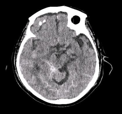
What is the abnormality on this image? What is a pertinent negative?
Moderately extensive acute subarachnoid blood products bilaterally, greatest burden centered within the perimesencephalic cisterns on the right.
No aneurysm identified. No intraparenchymal, intraventricular, subdural or extradural haemorrhage or collection.
What observational parameters would you want to observe someone within?
Arterial line to keep systolic BP 110-140mmHg.
You want to maintain cerebral perfusion (hypotension SBP <100mmHg causes ischaemia) and prevent re-bleeding (SBP >150mmHg associated with re-bleeding).
What is the sensitivity of non-contrast CTB to exclude this diagnosis within 6 hours of onset of symptoms?
100%.
It is slightly more difficult to pick up if it is a small bleed, a posterior fossa bleed, or the patient has low haematocrit (as this decreases the attenuation of blood).
ECG of the Week
62yo male presents with chest pain while in the gym at 10am.
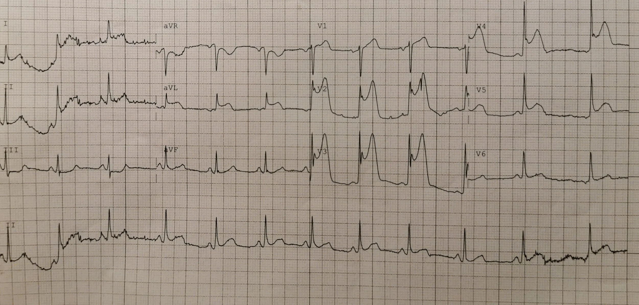
Describe the ECG.
Normal sinus rhythm showing anterior and lateral STEMI with reciprocal depression in Lead III.
His chest pain resolves in Resus. The following ECG was then taken.
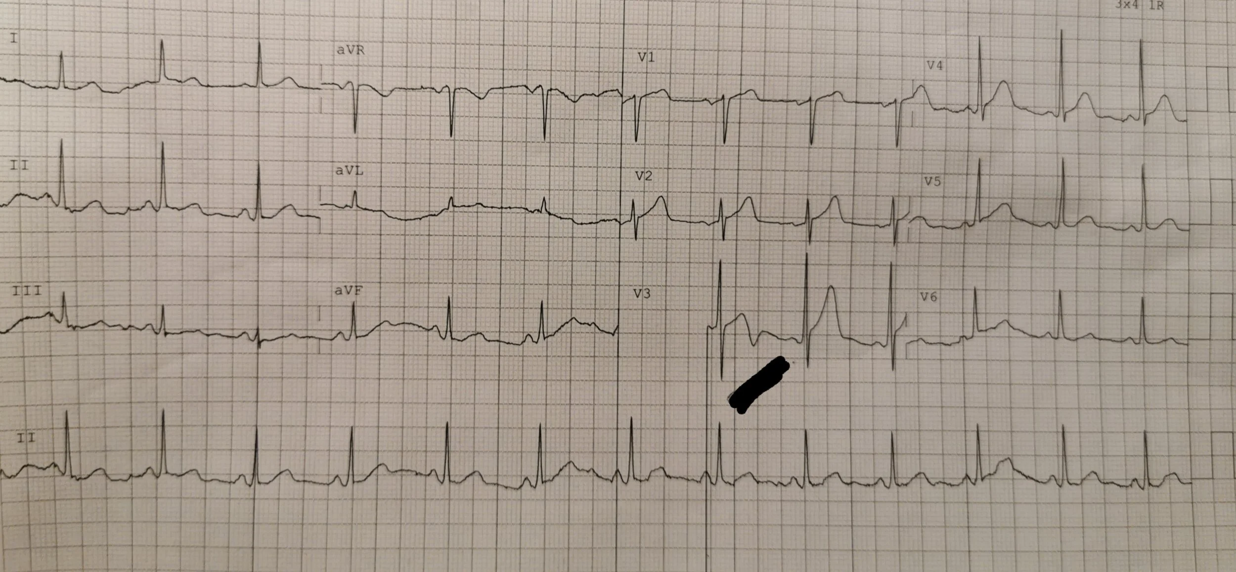
Describe the ECG.
Normal sinus rhythm with raised ST segments in V1-4 (looks more like benign early repolarisation with S-waves dipping below PQ interval in V2 and V3) and a biphasic T wave in V3.
What is the ECG wave above the black mark suspicious for? (Particularly in a pain-free ECG.)
Wellens’ syndrome (Type A with biphasic T waves in the anterior leads). This needs to be recognised because it represents critical proximal LAD stenosis and will cause anterior MI within 9 days (if not dealt to by PCI or CABG).
Note: The biphasic T wave changes of Wellens’ syndrome only show up on a pain-free ECG. Classically during pain, there is paradoxical pseudonormalisation of ST segments. (Which wasn’t the case with this patient, although he did have an LAD stenosis stented.)