24 February 2021 - Cardiovascular I
Program
1000-1130 - Morbidity & Mortality Meeting - Kirsten Cassidy
1130-1155 - Pacing - Matt Bray
1155-1203 - ECG of the Week - Sarah Bannerman
1203-1210 - Image of the Week - Ben Lee
1210-1230 - How to Give a Talk - Alex Handrinos
1230-1300 - Lunch
1300-1445 - Small group practical sessions (45min each)
Cardiac simulation
Pacing and Defibrillation
POCUS Echo
ABGs
Separate program for OSCE candidates as arranged with Brendan Morrissey.
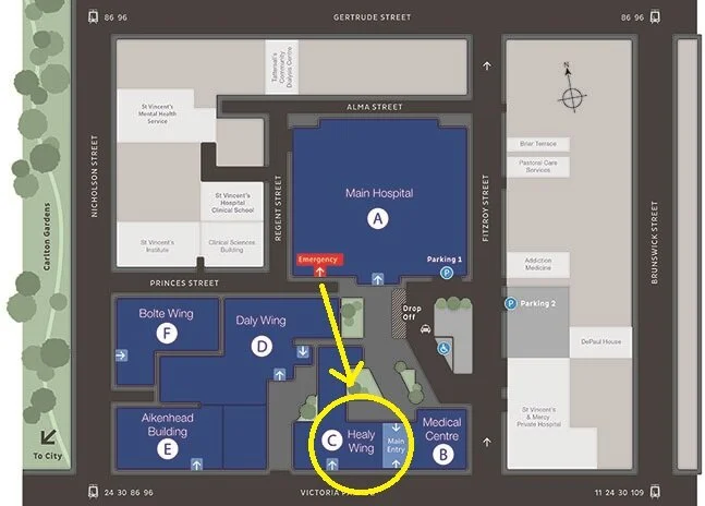
Teaching this week will be held in the Education Centre on Level 3 of Building C (Healy Wing).
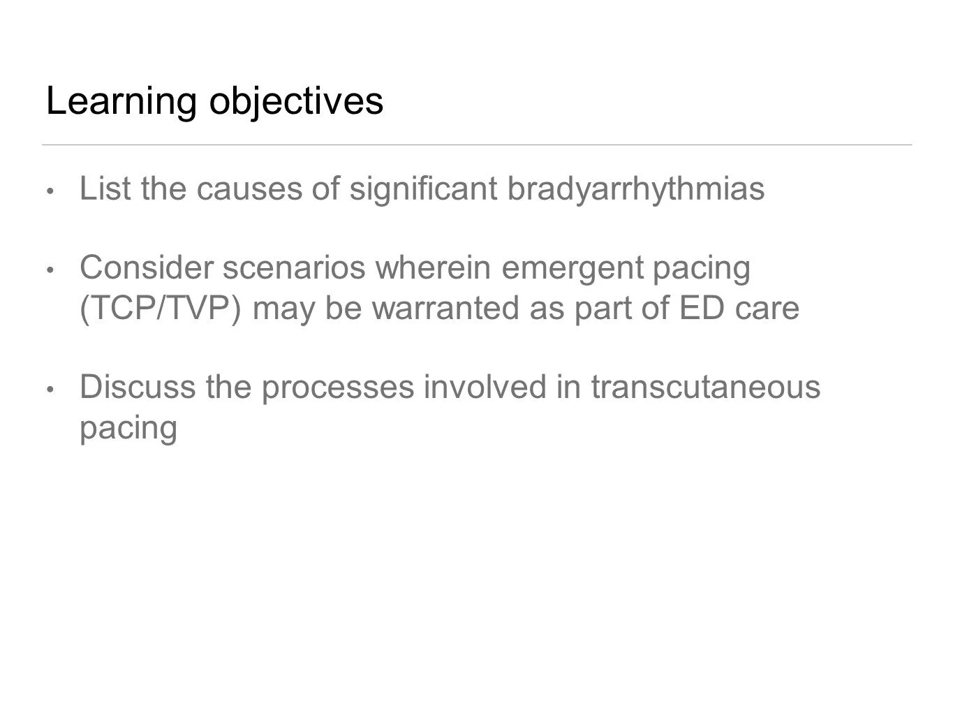
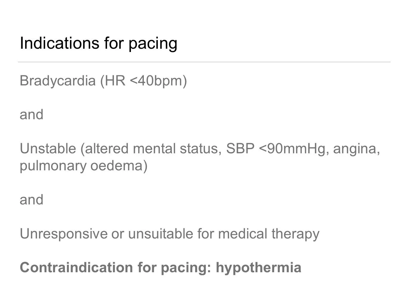
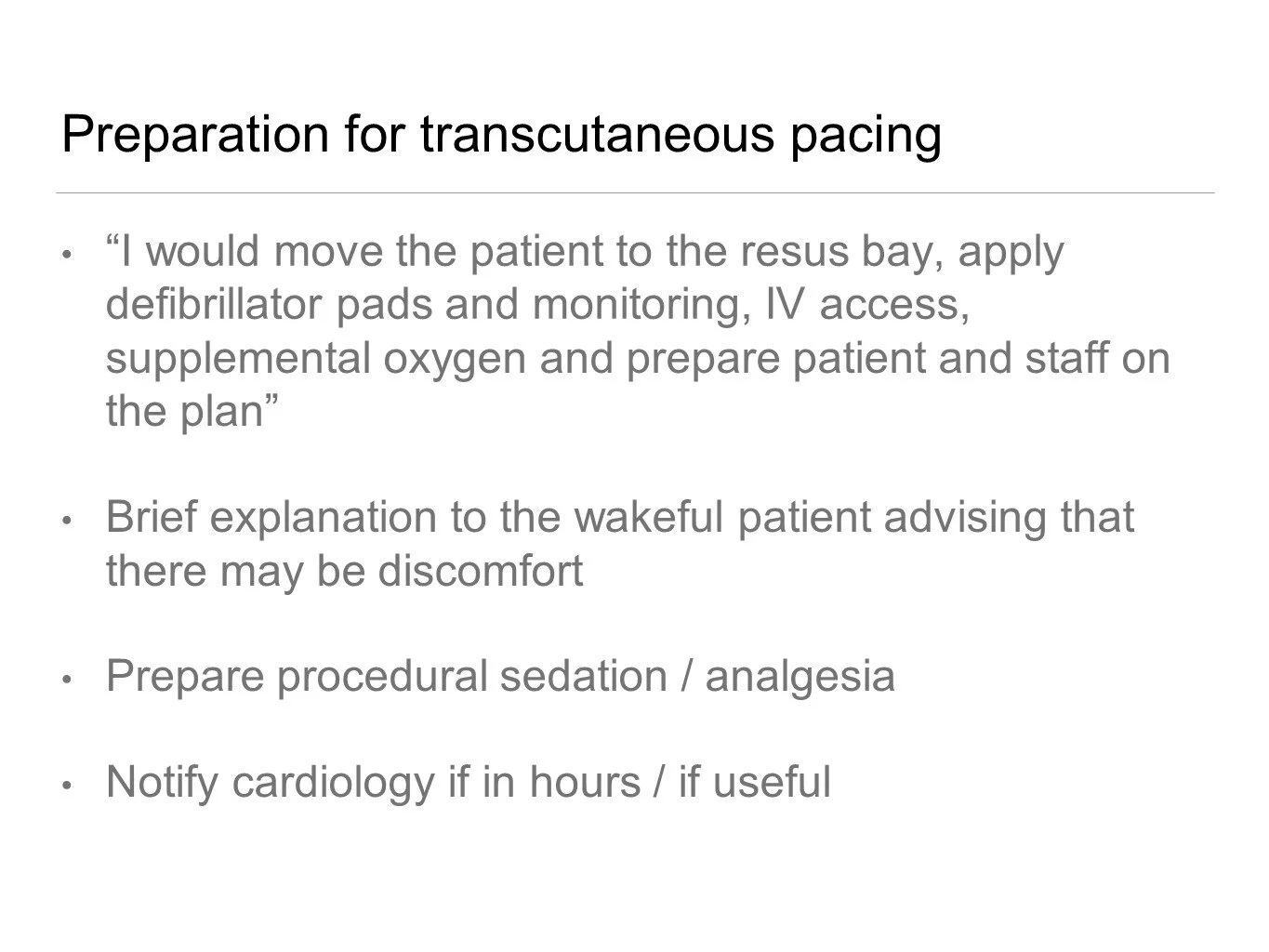
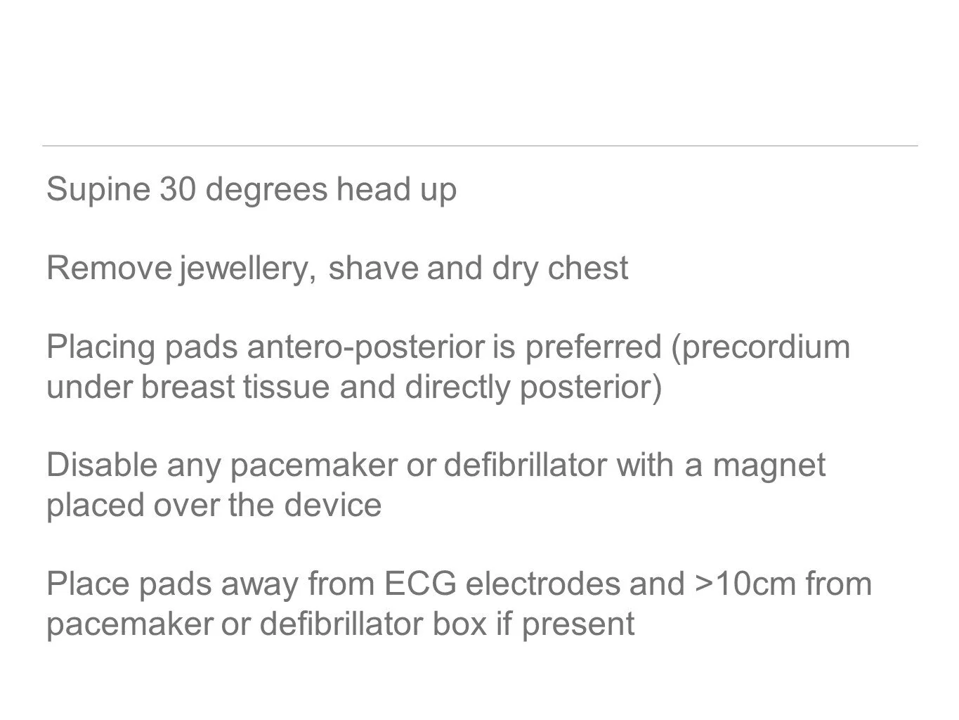
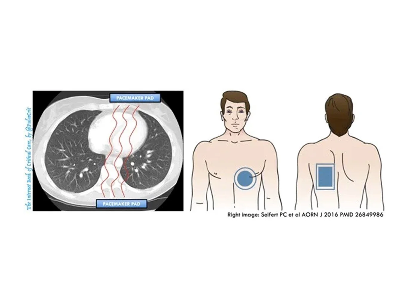
Matt Bray’s Pacing Talk
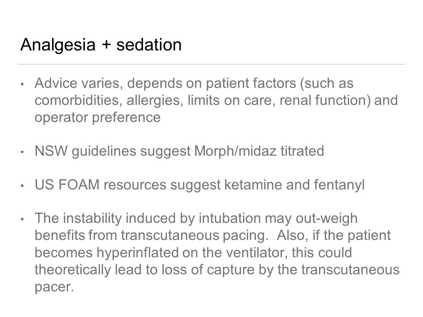
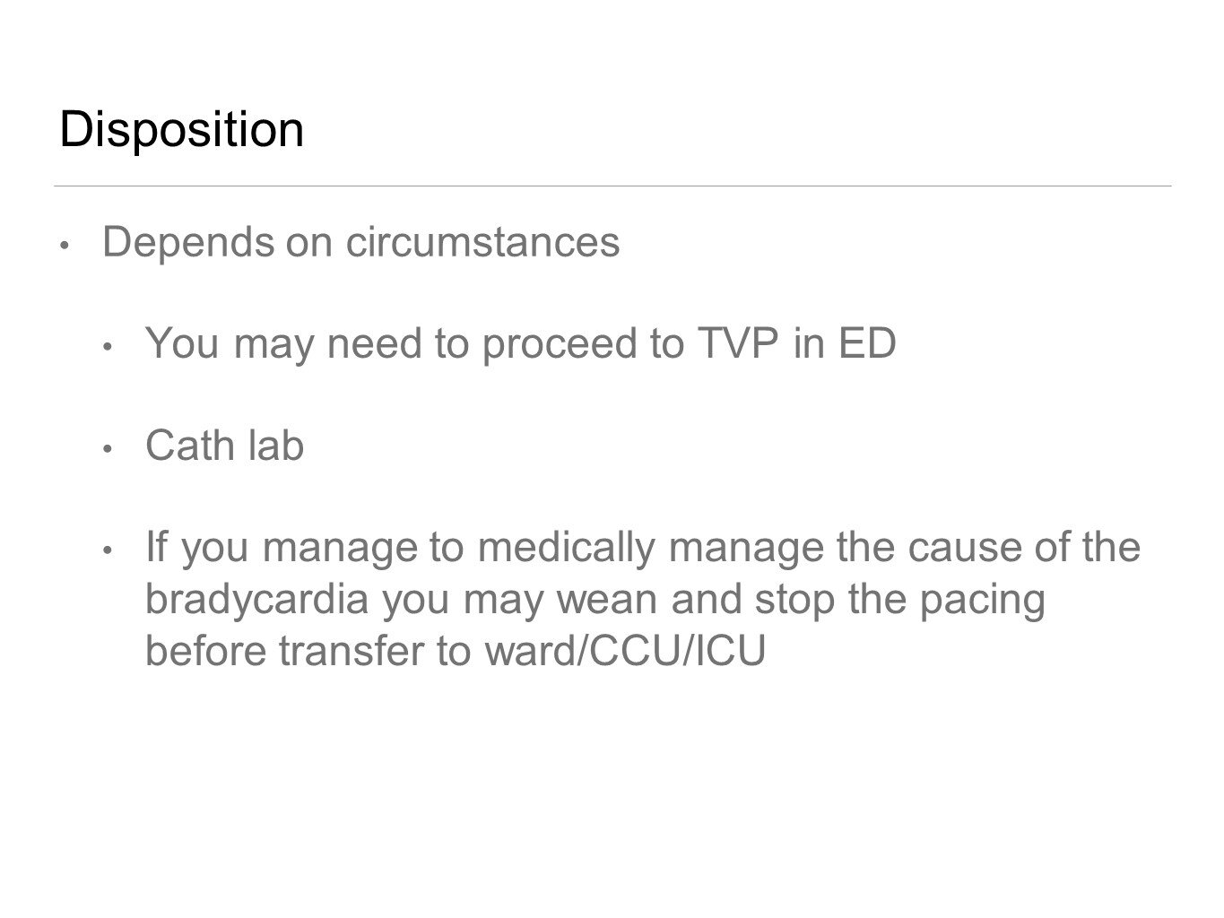
How to float a pacing wire:
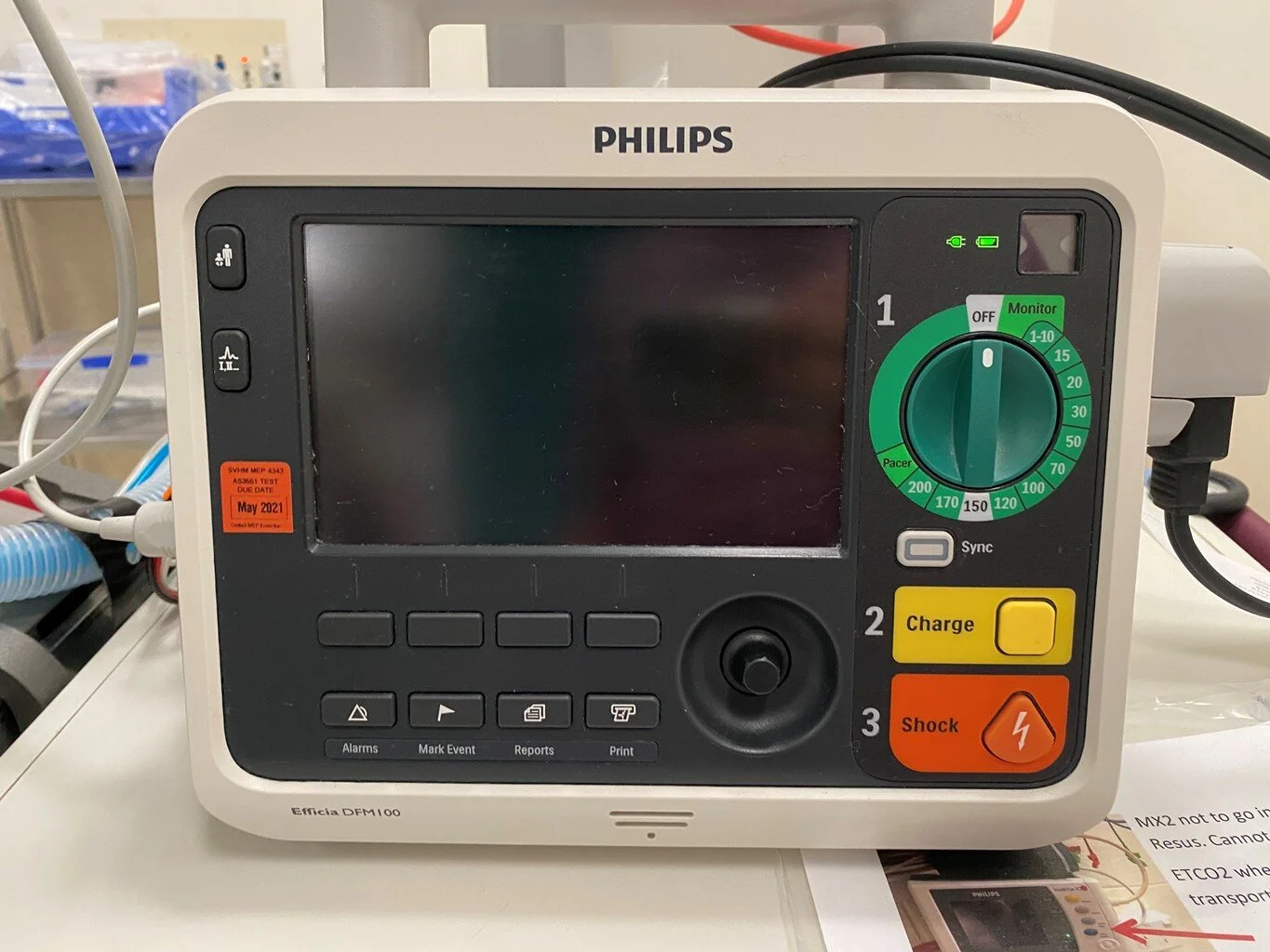
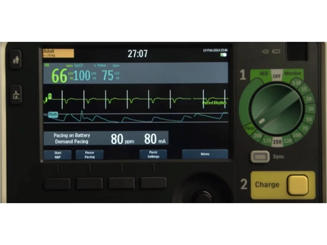
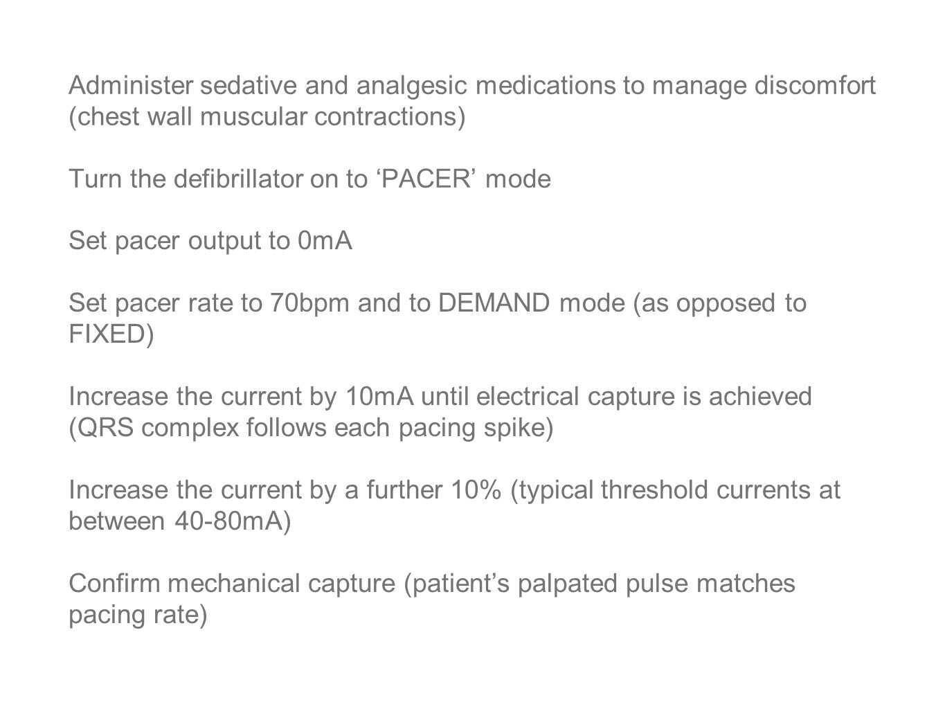
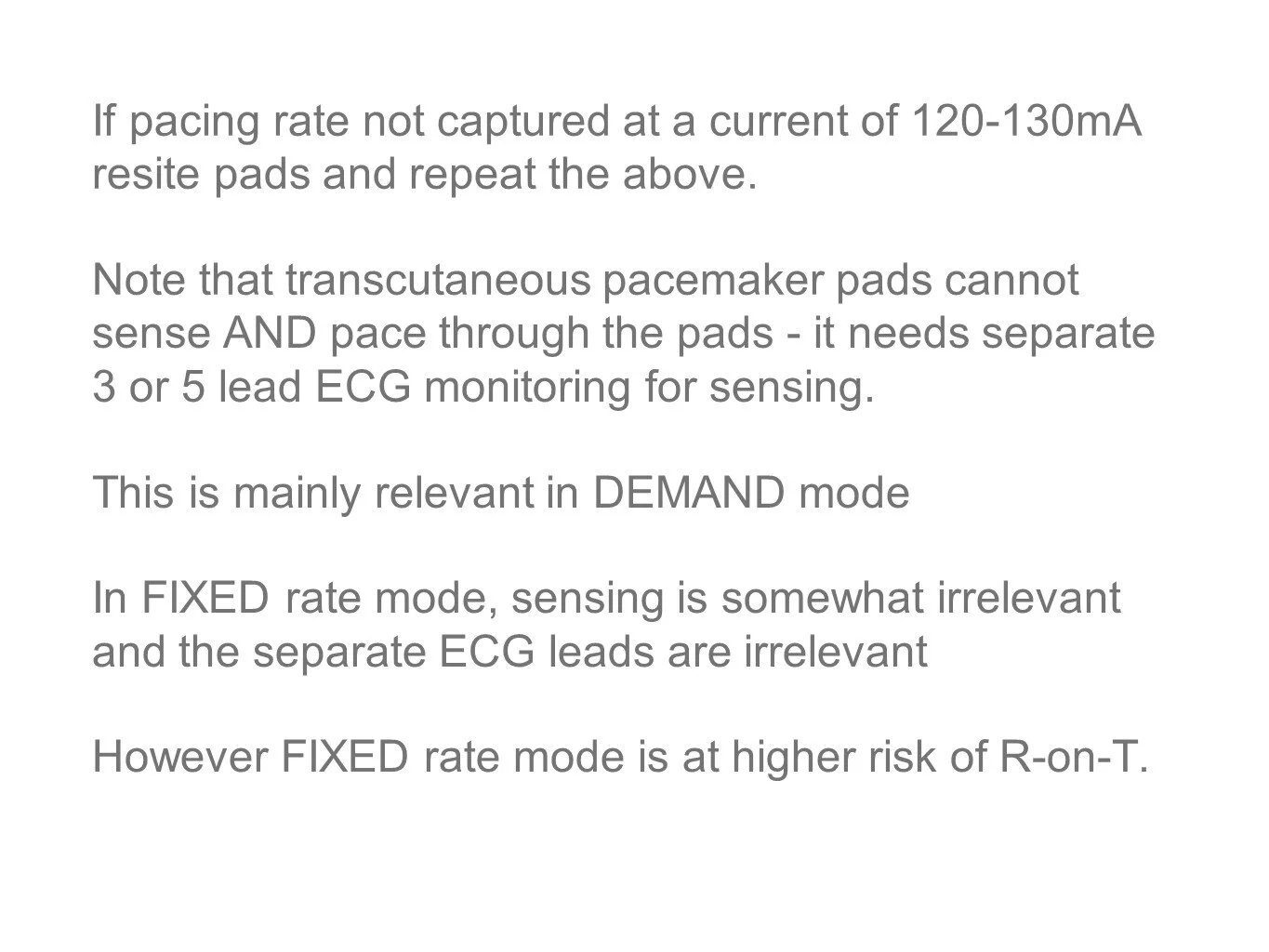
How to use the pacing function on our ED defib machines:
Key Learning Points
Thanks to Gem Ridley for this week’s Teaching Pearls:
Morbidity & Mortality Meeting
If you have clinical suspicion of a Lisfranc fracture, ensure you emphasise this to Radiology when requesting a CT
Be aware of the speed of bikes on triage notes. At the record-winning event, Usain Bolt's average ground speed was 37.58km/h = Cyclists are therefore always travelling slower than Usain at 20km/h.
Ben Lee’s Neuro Exam
Extend wrist - Radial
Abduct fingers - Ulnar
Circle of “OK” - Median
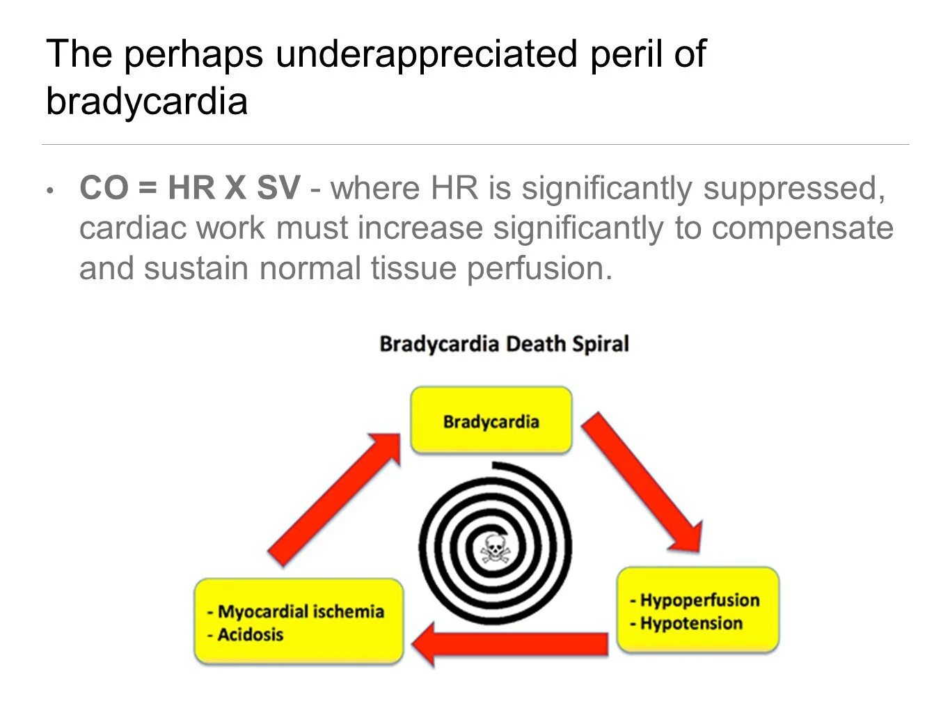
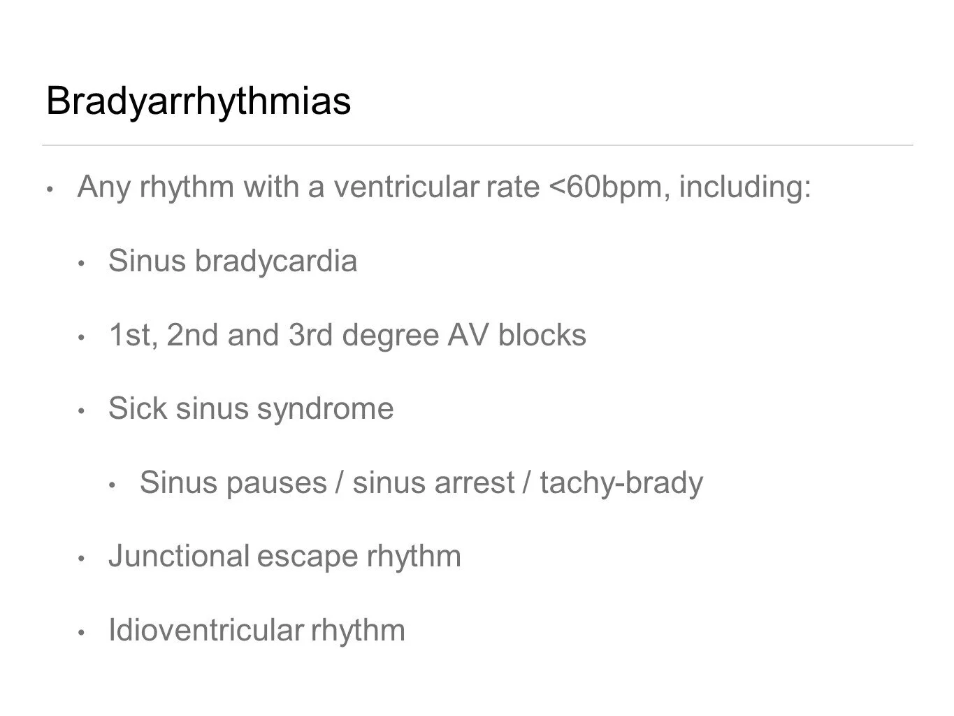
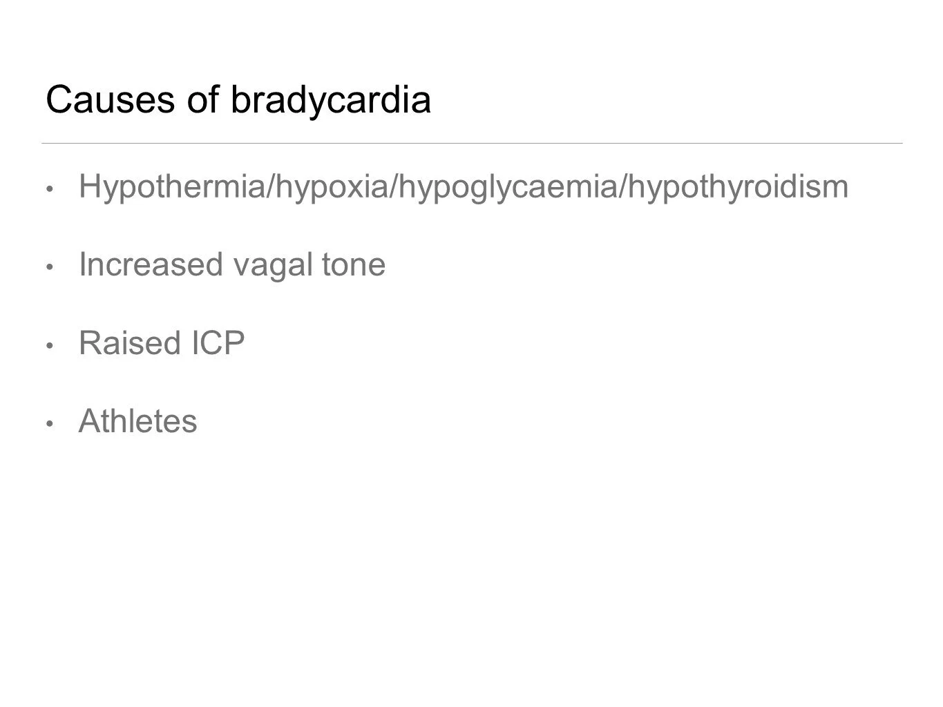
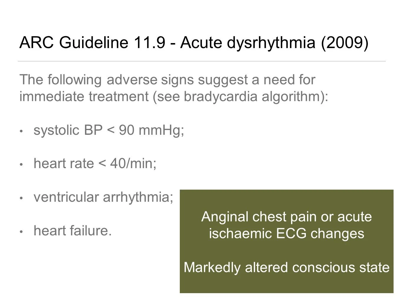
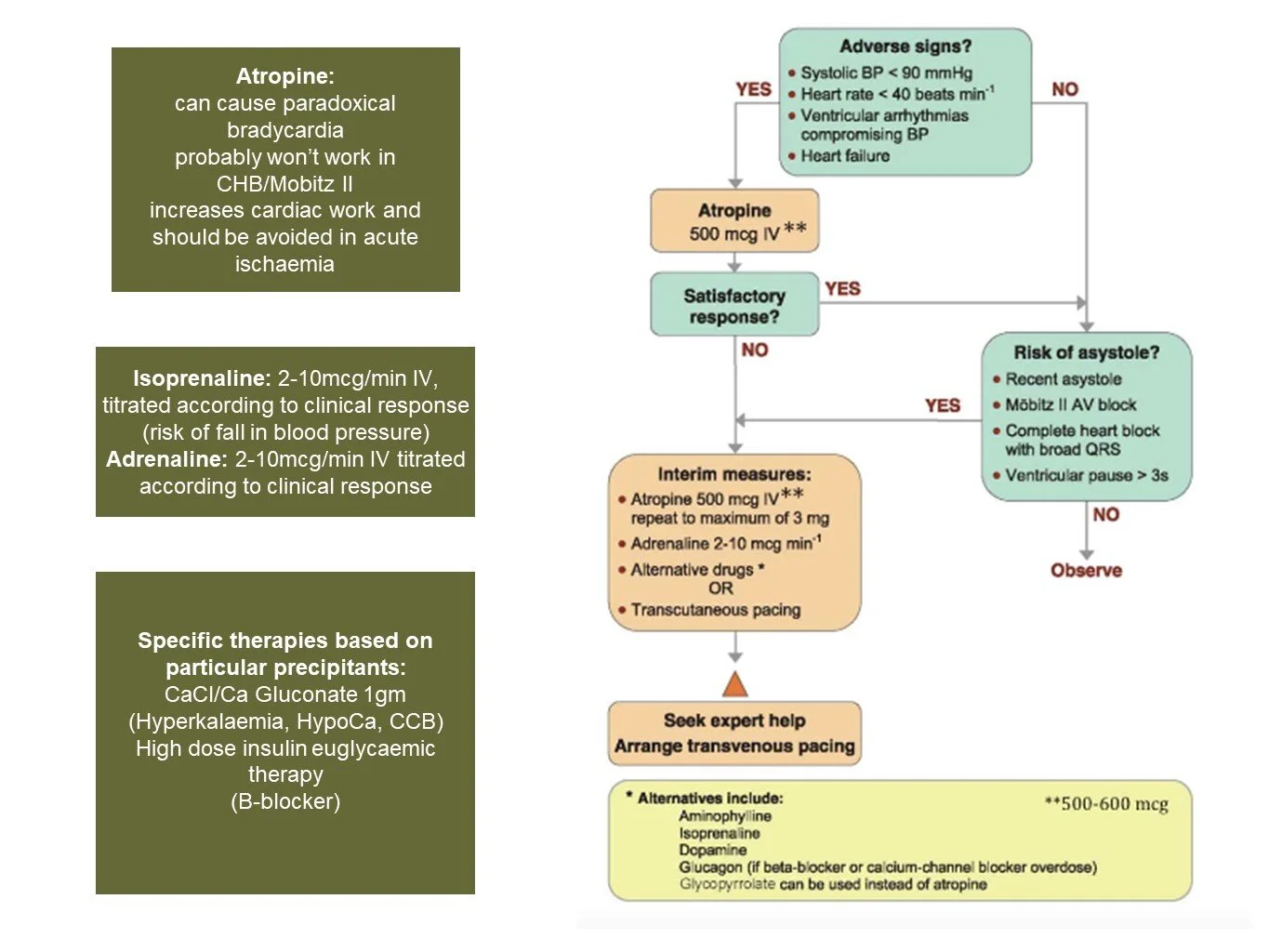
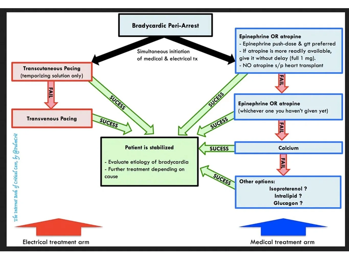
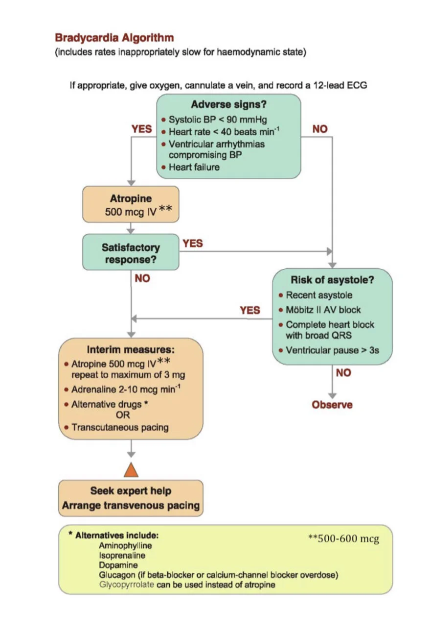
Bradycardia
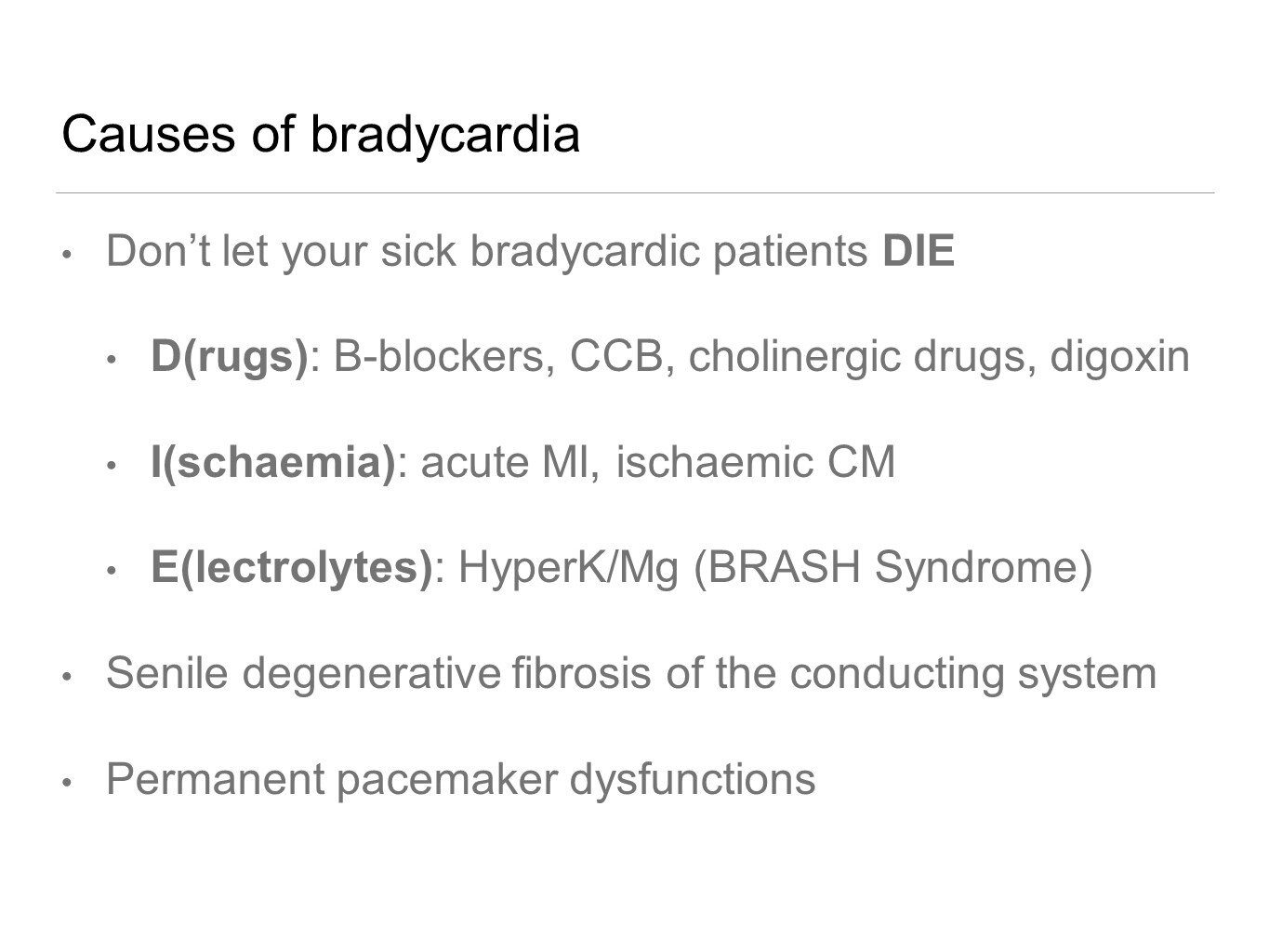
Causes — “Don’t let your patient DIE”
Drugs — beta blocker, calcium channel blocker, cholinergic, digoxin
Ischaemia — acute myocardial infarction / ischaemic cardiomyopathy
Electrolytes — BRASH for bradycardia, renal failure, AV node, shock, hyperkalaemia
Transcutaneous pacing — ensure you have your 3-lead attached to the defib as well as the pads!
Australian Resuscitation Council’s guidelines to managing acute dysrhythmias here
ECG of the Week
An asymptomatic 84 year old man is referred by his GP to ED for review of an incidental finding of bradycardia.
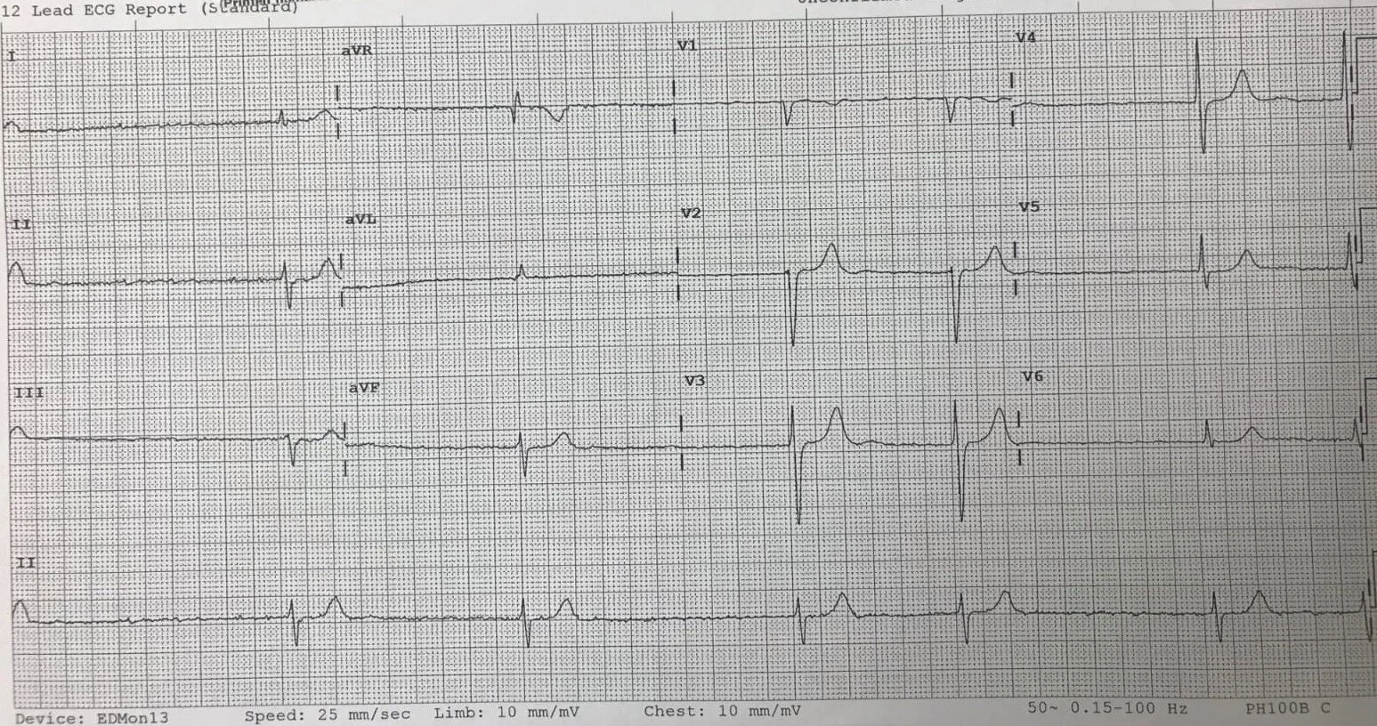
List three abnormalities.
● Bradycardia - 36 (30 - 50)
● Irregular
● No p waves
● Left axis deviation
This is slow AF, i.e. AF with a slow ventricular response. Important normalities to note are a narrow QRS and no T wave inversion.
List causes of this presentation.
Medications - beta blockers*, digoxin*, calcium channel blockers, amiodarone, clonidine, organophosphate toxicity
Cardiac (heart blocks including complete) - ischaemia, myocarditis, cardiomyopathy, congenital, sick sinus syndrome*
Hypoxia
Electrolyte abnormalities (high K and low Mg)
Hypothermia (< 35 °C)
Raised ICP (Cushing’s triad)
Hypothyroidism
Autoimmune
Sarcoidosis/Amyloidosis
Physiological (extreme fitness, vagal tone)
*Class IIb evidence in asymptomatic patients
What (definitive) management should be considered?
Withhold medications
Address reversible causes (e.g. high K or low Mg, hypothyroidism, etc)
PPM (Class IIb evidence in asymptomatic patients)
Image of the Week
29 year old lady presents having been assaulted with a fall to the floor and head strike.
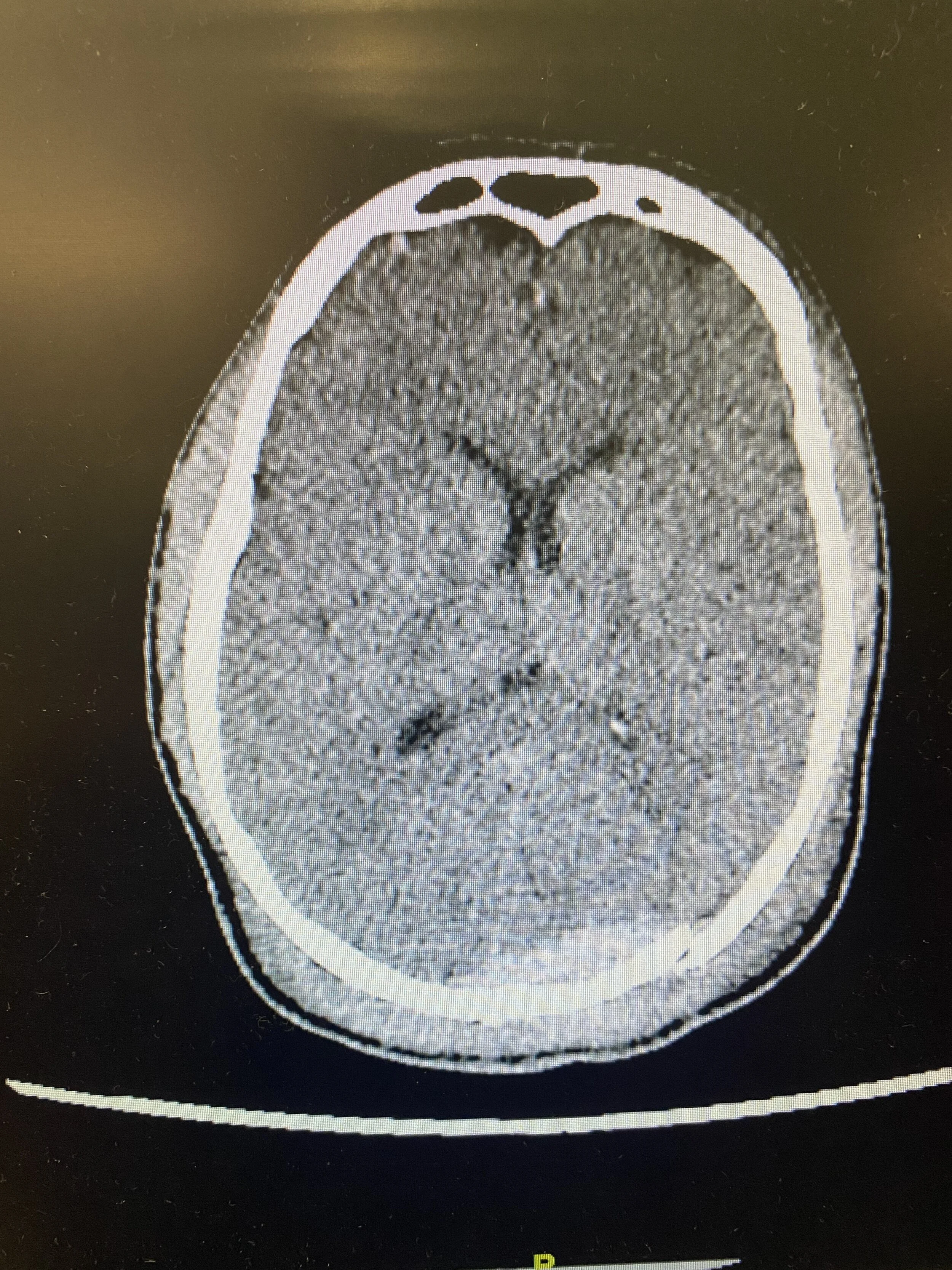
Name 4 pertinent traumatic finding on this CT brain image.
Large occipital swelling
Occipital fracture
10mm acute subdural haematoma
Contrecoup injury to right frontal lobe
ADDIT: Hey guys, apologies for the quality of the CTB slice. It was taken off a phone so some of the abnormalities are difficult to see. (It’s not you, it’s the image!)
Name 2 pertinent negatives of this CT scan which you may report to the Neurosurgical Registrar.
No local mass effect/effacement of ventricles
No midline shift
Name 3 spaces/ventricles of the brain that can be seen on this image.
Frontal horn of lateral ventricles
Third ventricle
Choroid plexus