Program
1000-1100: Ask the expert, Q&A: Nirupa Sachitananda (endocrinologist)
1100-1130: Jannet: Thyroid emergencies, with coffee, give Sarah your orders
1130-1200: Hamed: Journal Club
1200-1230: Lunch
1230-1300: Jason: Adrenal insufficiency
1300-1320: Homelessness: Jennie Hutton
1320-1350: Recap, ECG and image of the week
1350-1400: Ben's case
1320 - OSCE and FEx groups with Brendan and Alex
Image of the Week
25M presents with R shoulder pain and reduced ROM post fall whilst intoxicated. Also reports HS with unknown LOC.
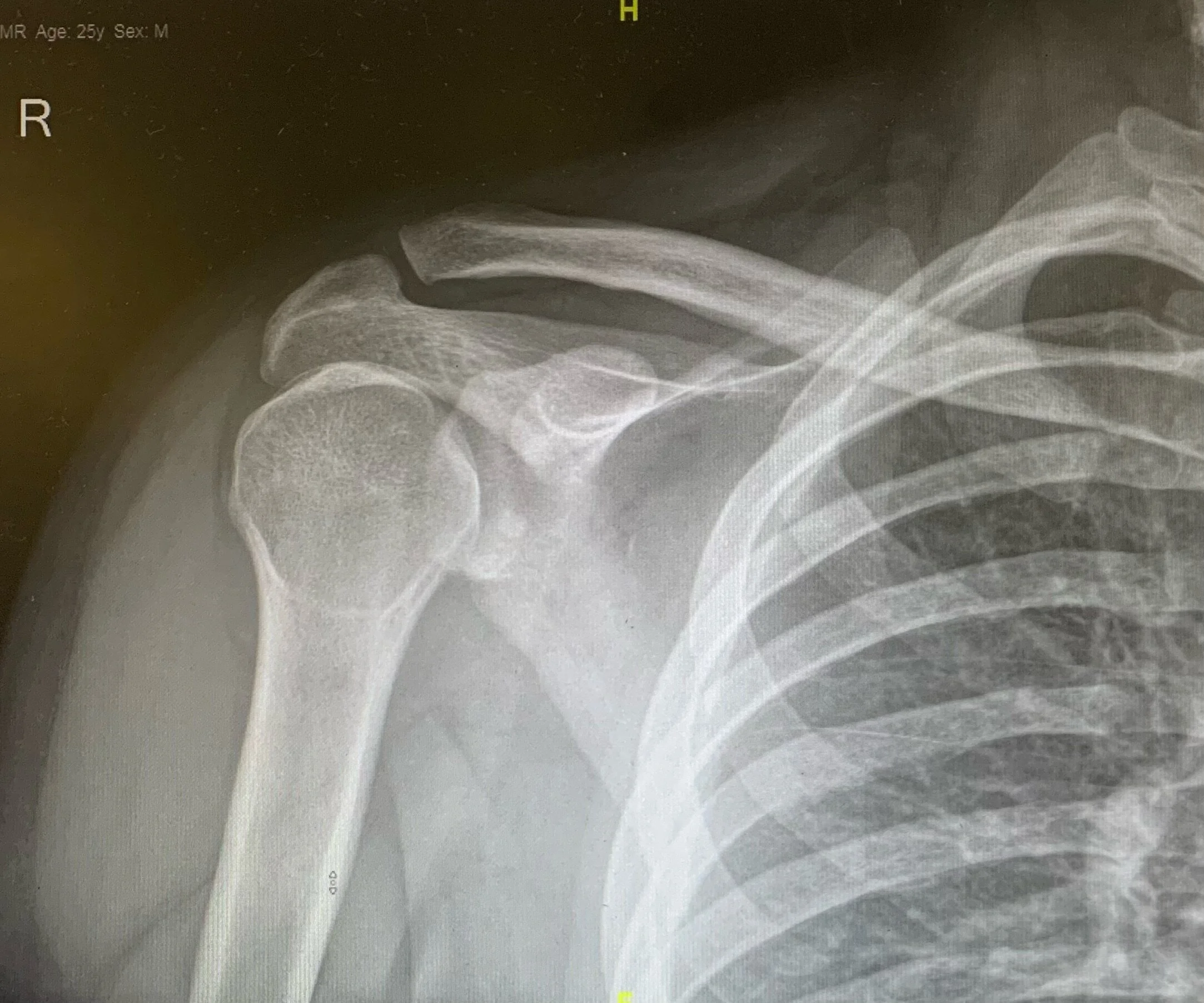
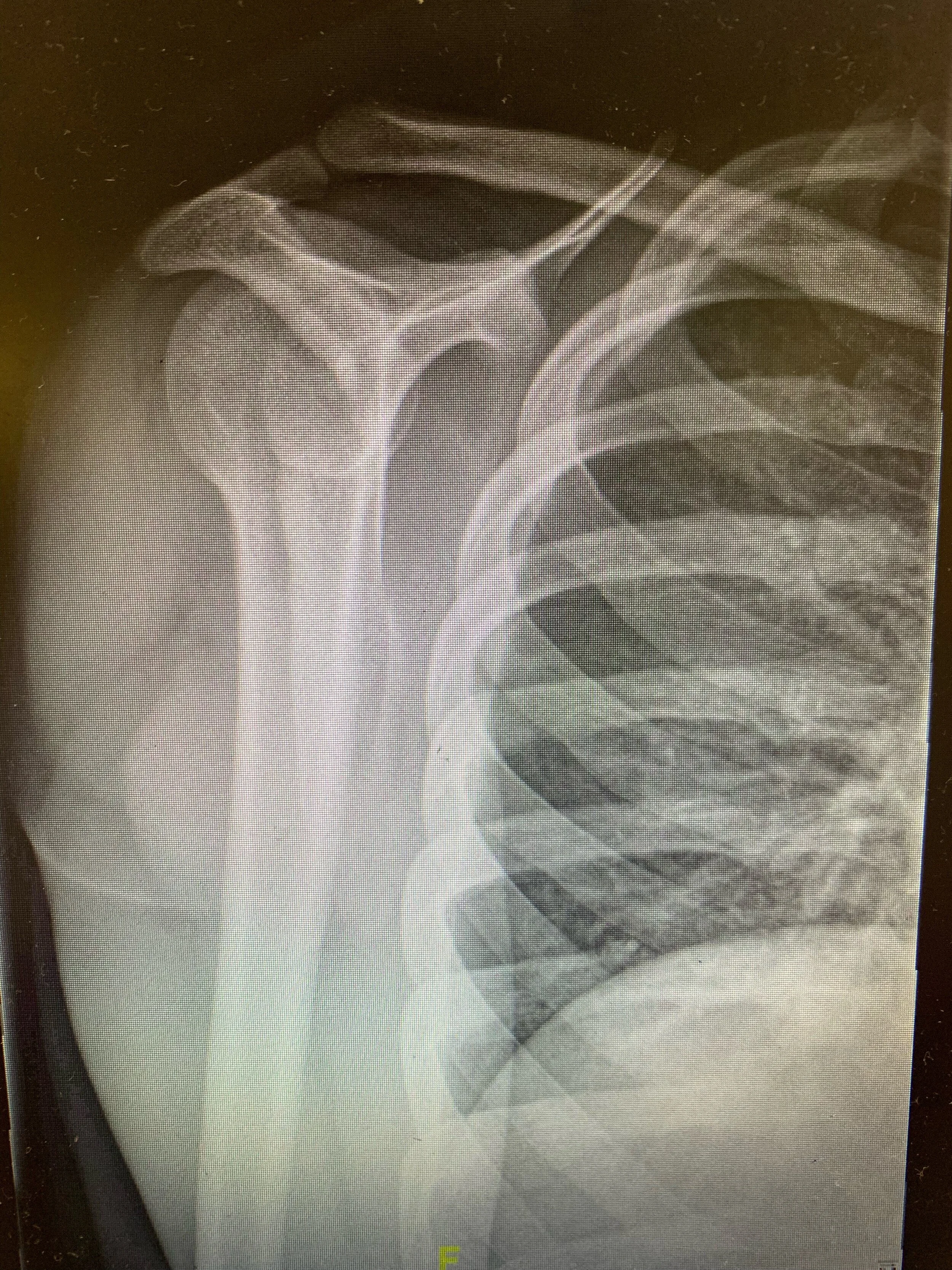
1) Describe the X-rays
Light bulb sign — the humeral head is internally rotated and therefore in the same axis as the shaft, resembling a “light bulb”. This is suspicious for posterior shoulder dislocation.
Other signs of posterior shoulder dislocation can be:
Trough line sign — two nearly parallel lines in the superomedial aspect of the humeral head
Loss of normal half-moon overlap sign — the glenoid fossa appears vacant due to lateral displacement of the humeral head
Rim sign — widened glenohumeral joint > 6 mm
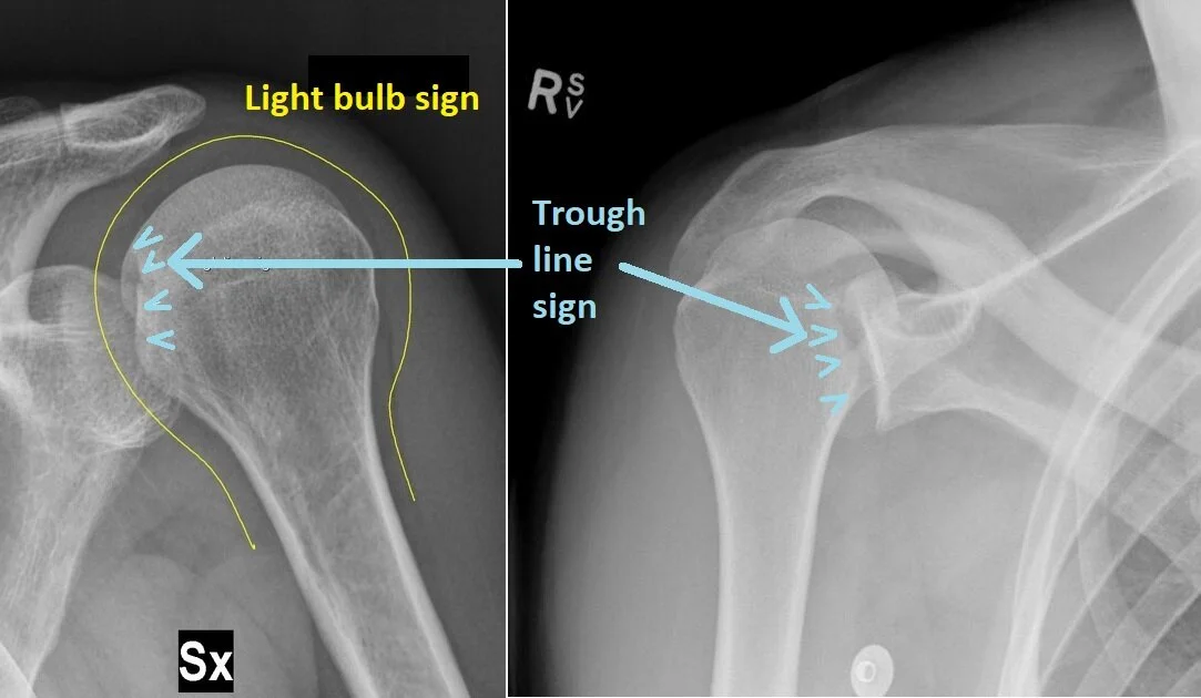
Light bulb sign — the humeral head is internally rotated, resembling a “light bulb”
Trough line sign — two nearly parallel lines in the superomedial aspect of the humeral head
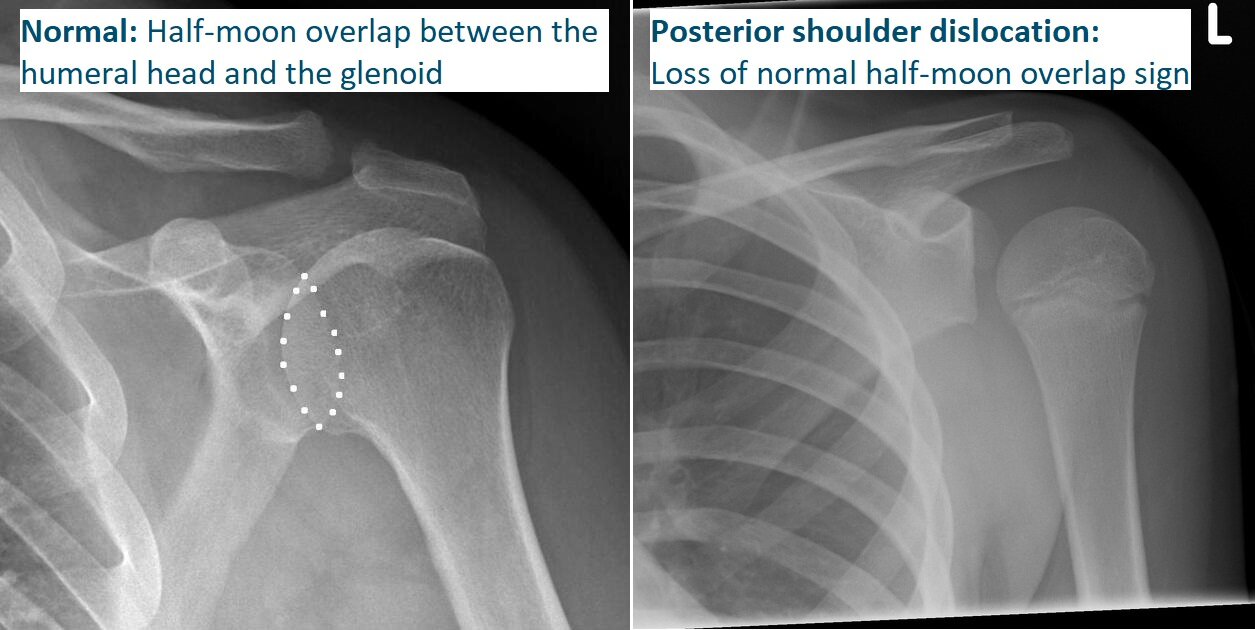
Loss of normal half-moon overlap sign — the glenoid fossa appears vacant due to lateral displacement of the humeral head
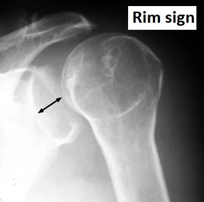
Rim sign — widened glenohumeral joint > 6 mm
Beware! The AP view can look normal in 50% of posterior shoulder dislocations. The scapular Y view has also been shown to be unreliable for diagnosing posterior shoulder dislocations.
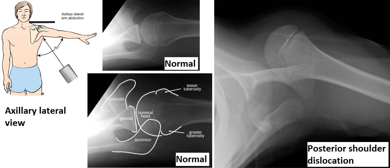
The best view to see a posterior dislocation is the axillary lateral view (this may be difficult for the patient to achieve, though).
2) What important positive or negative findings would you to document in terms of your examination?
Neurovascular status
3) What are the priorities of your management of this patient in ED?
Analgesia
C-spine immobilisation
Primary and secondary survey
CTB and C-spine
Procedural sedation to reduce shoulder, e.g. using a reverse modified Kocher’s manoeuver