
14 April 2021 - Toxicology
Program
Teaching will be on Zoom this week:
https://zoom.us/j/5981463478?pwd=RThaZzNkL2w5bldBMlNHeE1zZFFaZz09
Meeting ID: 598 146 3478
Passcode: 2021
1000-1015 — Welcome & Recap — Jannet Chan
1015-1025 — ECG of the Week — Jason Aw
1025-1030 — Image of the Week — Ben Lee
1030-1130 — Addiction Medicine Q&A — Dr Martyn Lloyd-Jones, SVHM Department of Addiction Medicine
1130-1200 – Break
1200-1230 – ‘Mushroom Ingestion, from Magic Mania to Major Problem’ – Alasdair Williamson
1230-1300 – Serotonin Toxicity – Jacqueline Maplesden
Break-out group on Zoom:
1300-1400 – Teaching on the Run with Brendan Morrissey
1300-1500 — Primary Exam Group with Alex Swain (potentially may also be on site?)
On site:
1315–1445 Fellowship Written Exam Group with Alex Handrinos — Jannet Chan & Ezra Lim. Breakout room 2, Education Centre
1330-1500 – Ultrasound with Mark Rugless — Ben Lee & Jess Robinson
ECG of the Week
34yo lady who is 36/40 pregnant presents with nausea, vomiting and left-sided chest discomfort. She has been vomiting for 12 hours. Her systolic BP is 95mmHg.
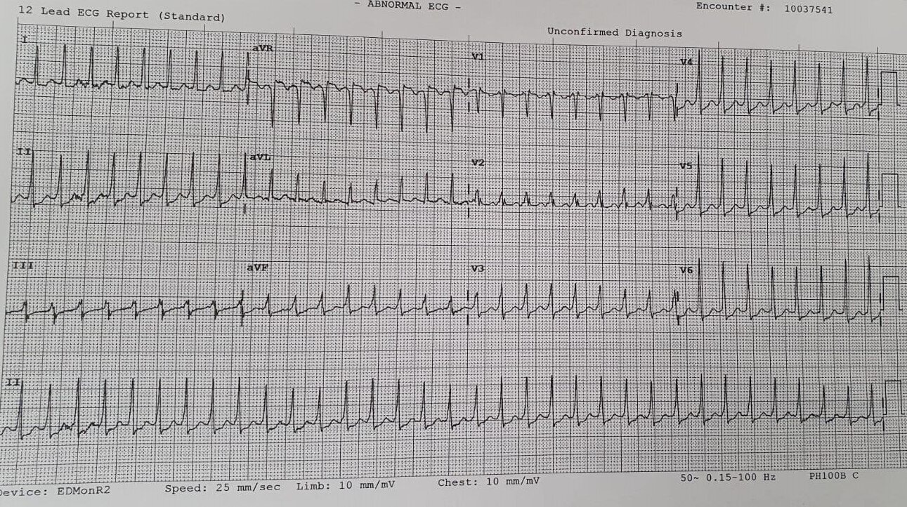
1) Describe the ECG
Narrow complex tachycardia, i.e. SVT
2) What would be your initial management?
Move to Resus cubicle
IV fluids
Correct electrolytes
Call O&G
Attempt Valsalva manouevre (there’s a new kid on the block if the modified Valsalva doesn’t work — it’s called the reverse vagal maneouvre)
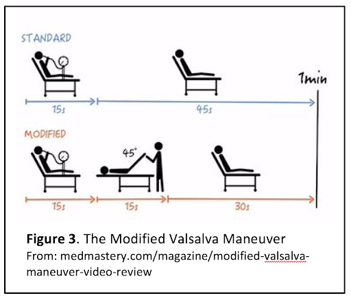
Standard and Modified Valsalva manouevres
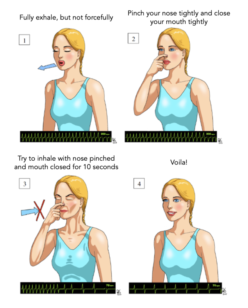
Reverse Valsalva manouevre
3) If this patient became hypotensive, what would you do?
Alert O&G and Paeds teams about the potential impending need for an emergency C-section
Call Cardiology for advice
Adenosine is the drug of choice => If that fails, DC cardiovert
Monitor the foetus throughout
Image of the Week
22yo lady presented with 3 days of SOB and dizziness. Vital signs are RR 16, SpO2 100%, HR 93.
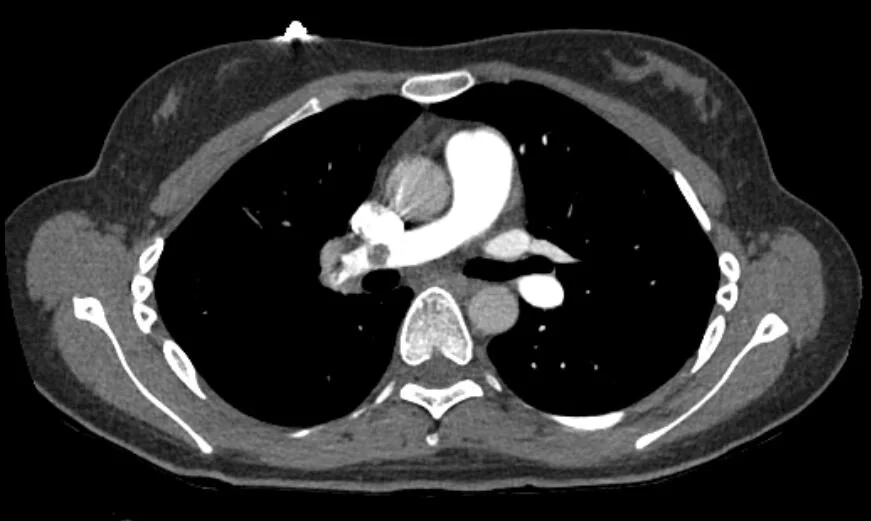
1) Describe the image and the abnormality seen
Axial view of CTPA showing the right pulmonary artery and part of the left pulmonary artery
Non-occlusive embolus in the right pulmonary artery
2) What possible findings may be seen on an ECG of this patient?
Nothing abnormal
Sinus tachycardia
Right axis deviation
RBBB
Anteroseptal ST elevation or depression
Anteroseptal T wave inversion
S1Q3T3
Likely PEA or asystole in cardiac arrest
3) What findings seen on TTE or CT may suggest right heart strain? Give 3 answers.
Abnormal position or flattening of interventricular septum
Right ventricular enlargement (bigger than left)
Pulmonary trunk enlargement (bigger than aorta)