28 April 2021 - Gastrointestinal
Program
1000-1030: Management of Upper GI Bleed (Medications) — Gemma Ridley
1030-1115: Breaking Bad News OSCE Session — Brendan Morrissey
1115-1145: Management of Upper GI Bleed (Procedural intervention) — Jannet Chan
1145-1215: Lunch
1215-1300: Gastro Sim — Alex Handrinos
1300-1400: Rapid Fire Gastro Updates — Robbie Little & Michael MacIsaac (Gastro Regs)
Approach to Acute Liver Failure
Approach to Decompensated Chronic Liver Disease
Acute Severe Ulcerative Colitis
1400-1415: Recap of past 2 weeks — Jason Aw
1415-1425: ECG of the Week — Sarah Bannerman
1425-1430: Image of the Week — Ben Lee
Teaching this week will begin in the Education Centre on Level 3 of Building C (Healy Wing).

Image of the Week
20yo lady presents with abdominal pain and vomiting for the past 12 hours. She has recently been commenced on prednisolone and erythromycin for a pimple on her eyebrow. She has a history of regional pain syndrome. She is currently hyperventilating with RR 32. The triage nurse sends her to Cubicle 14.
Here is her arterial blood gas:
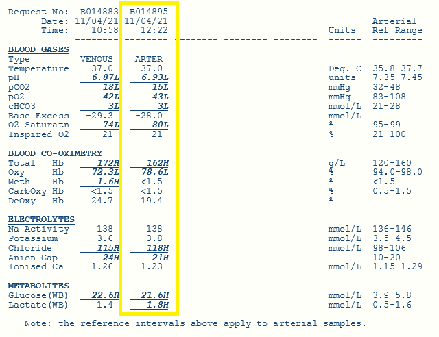
1) Please interpret the blood gas. Show your working where appropriate.
Patient has a profound metabolic acidosis. There is clear respiratory compensation (the CO2 is notably low).
Using the Fenkl-Stewart approach, we use base excess (BE) instead of HCO3 for our calculations. A negative base excess suggests that there is an excess of acid. In the Fenkl-Stewart approach, we compare the measured base excess (i.e. what the machine has told us) to our calculated base excess (i.e. based what we calculate).
This patient has a measured BE of -28. This means she has an excess of acid of 28.
BE -4, i.e. an excess of acid of ≥4, is considered metabolic acidosis
BE -10 to -20 indicates moderate metabolic acidosis
BE -20 indicates severe metabolic acidosis
Excessive acid can come from 4 sources:
Strong ion difference (SID)
Lactate
Albumin
Ketones
To figure out what’s causing the metabolic acidosis, you need to calculate how much of the base excess can be contributed to each of the 4 sources (i.e. what proportion of the excessive acid comes from each source).
First, let's calculate how much of the measured base excess in this patient (i.e. BE -28) can be contributed to the strong ion difference (SID).
Normal SID in a healthy person should be 42. However in this patient...
SID = (Na + K)- Cl
SID = (138 + 4)- 118
SID = 24
The difference between normal SID and our calculated SID = 42 - 24 = 18. This means that the SID accounts for an excess of acid of 18, i.e. BE -18.
If the strong ion difference was the only problem, the measured BE would be -18. But it's -28! So there is something else contributing to the base excess.
Second, let's calculate how much of the measured base excess can be contributed to lactate.
Normal lactate is 1. However in this patient, lactate is 1.8.
This means that lactate accounts for an excess of acid of 0.8, i.e. BE -0.8.
Third, let's calculate how much of the measured base excess can be contributed to albumin. For every drop in albumin of 4, the BE should increase by +1.
Unfortunately in this patient we don't have an albumin (it's not on the ABG!). However in most people, albumin would be normal. So for this patient, let's assume albumin is normal and doesn't contribute to the measured base excess.
So far, we have calculated that part of the measured excess of acid of 28 can be contributed to the SID (18) and lactate (0.8). However this still leaves us with an excess of acid of 9.2 that is unaccounted for. It must be ketones!
Lastly, let's correct her K for pH. When the pH drops (i.e. becomes more acidotic), K should move out of the cell into serum. That is, as the pH drops, your K should rise and be high. For every drop in pH of 0.1 below pH 7.4, K should rise by 0.5 above 5.0.
Given that her pH is 6.9, this means the pH has dropped 7.4-6.9 = 0.5. This is a drop of 0.1 x 5 = 0.5.
Therefore we expect her K should rise by 0.5 x 0.5 above 5.0. That is, it should be 0.25 above 5.0. Therefore the expected K is 5.25.
Her calculated K should be 5.25, however her measured K is 3.8. This means she is very hypokalaemic!
2) What is your diagnosis?
DKA
3) How would you manage this patient?
DKA pathway
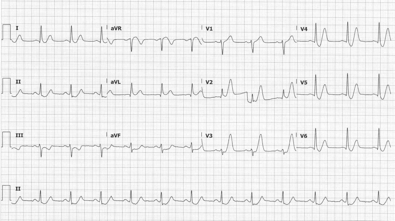
ECG of the Week
67yo lady presents with dizziness. On further questioning, she describes “pressure’ in her chest. Her only past medical history is hypertension. This is her ECG: