
7 April 2021 - Respiratory II
Program
1000-1015: Recap & quiz of last week’s teaching — Phil Ward
1015-1020: Image of the Week — Ben Sinnett
1020-1030: ECG of the Week — Gemma Ridley
1030-1130: Respiratory Controversies — Matt Conron (Respiratory Physician)
1130-1200: Break
1200-1330: ED/ICU Case Discussions
1330-1400: Lunch
1400-1430: Ventilator Workshop — Alex Handrinos
NOTE: Alex Handrinos has uploaded some excellent videos on ventilation — please have a look before teaching :)
Teaching this week will begin in the Education Centre on Level 3 of Building C (Healy Wing).
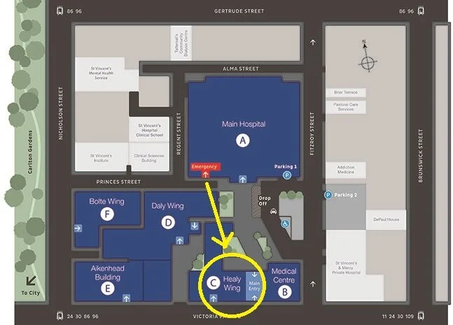
Image of the Week
75F transferred from a nursing home with a one day history of abdominal pain, distension and vomiting. Past history includes renal impairment and dementia.
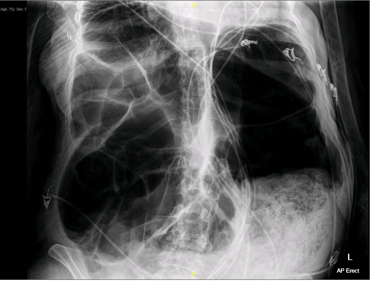
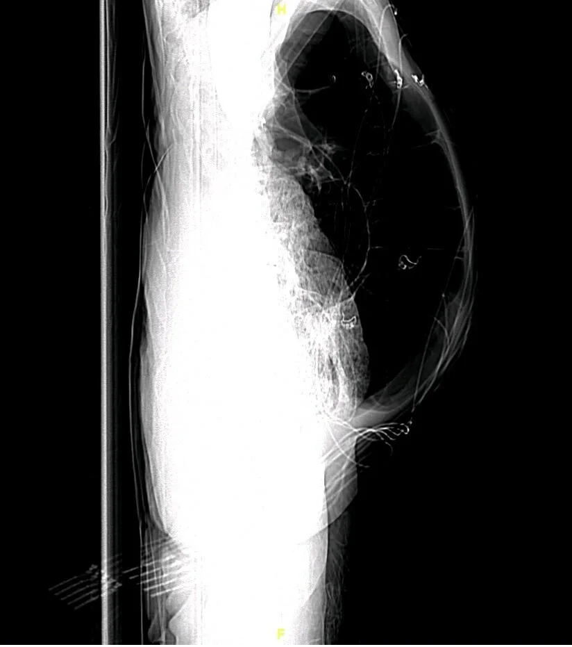
1) Identify 3 abnormalities on the plain film.
Significantly dilated bowel (colon >6cm)
Large amount of faeces distally
2) Suggest 4 differentials, based on the AXR.
Large bowel obstruction
Toxic megacolon
Colonic pseudo-obstruction (Ogilvie’s syndrome)
Ischaemic colitis
3) On examination, the patient has active bowel sounds, while on the inevitable CT, no transition point is identified. What management options are available to you?
Treatment of Ogilvie’s syndrome:
Correct electrolyte imbalance
Cease opioids, calcium channel blockers, antidepressants, and antispasmodics
Nil by mouth, NGT insertion, bowel rest
Prokinetics, e.g. IV metoclopramide 10mg TDS, PO erythromycin 500mg TDS, IV neostigmine 2mg
4) Identify 2 life-threatening complications, if left unchecked.
Perforation with abdominal sepsis
Abdominal compartment syndrome
ECG of the Week
50yo man presented to ED with palpitations.
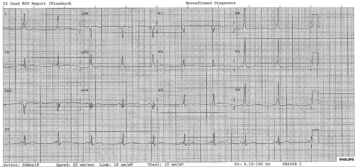
1) Describe the ECG.
Sinus rhythm with normal axis. Short PR interval. Slurred upstroke to QRS, most prominent in V4-V6. Isoelectric ST segments.
2) What syndrome is associated with these abnormalities. (Bonus point for types.)
WPW
Type A — tall R in V1
Type B — deep S in V1
3) What rhythm is this abnormality associated with?
Atrial flutter