25 August 2021 - Ophthalmology & ENT
Teaching this week will be on Microsoft Teams:
Program
1000—1020: Recap (Sunshine PEM and Haem week) — Andrew West
1020--1030: ECG of the Week — Luke Bayley
1030—1115: ENT Radiology — Leon Winata
1115—1130: Stretch & Coffee
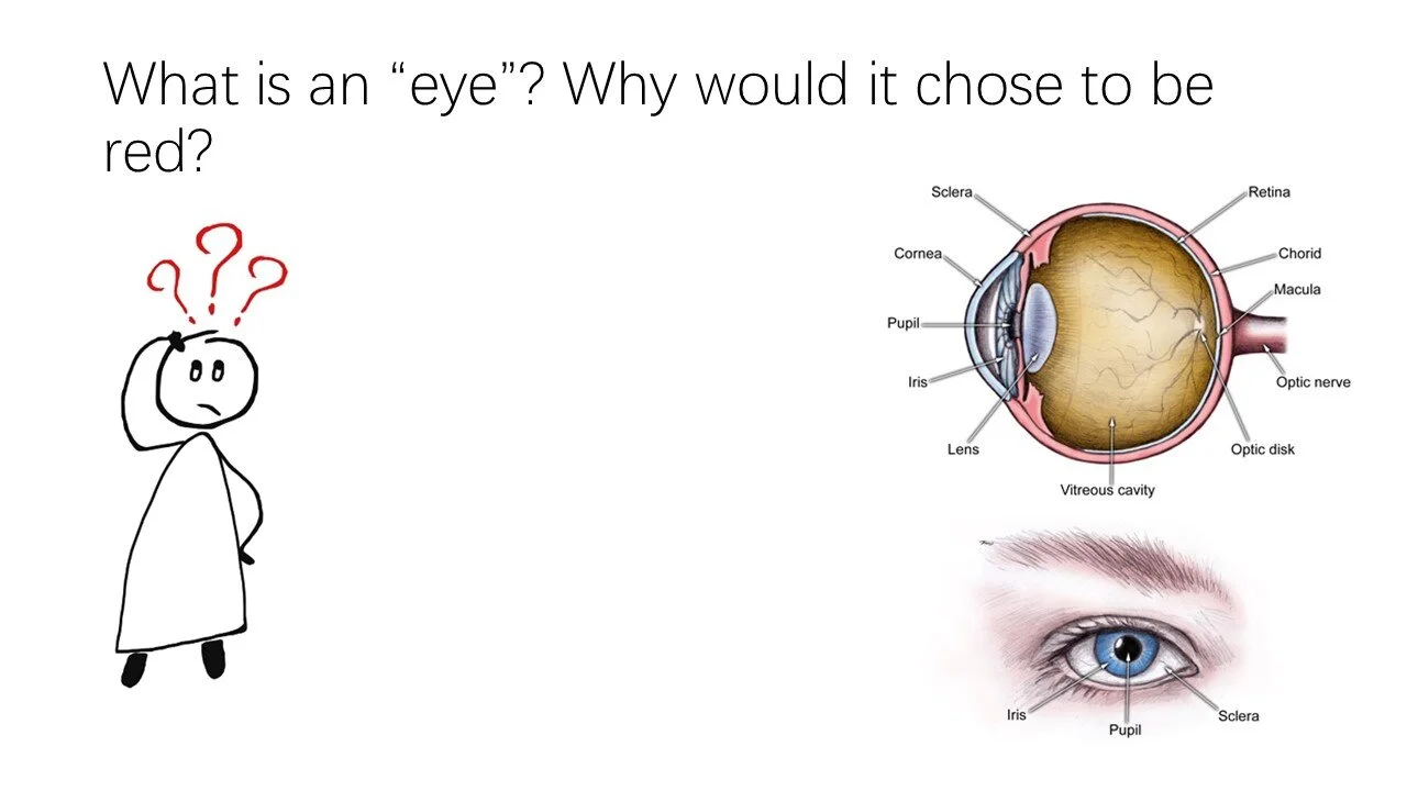
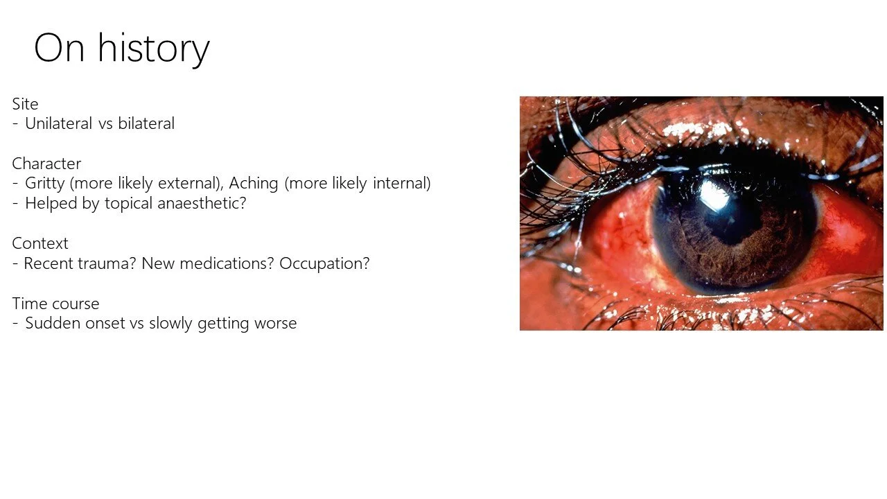
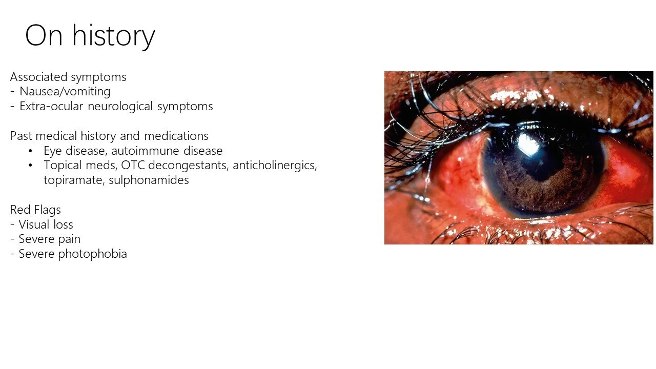
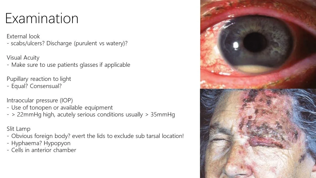
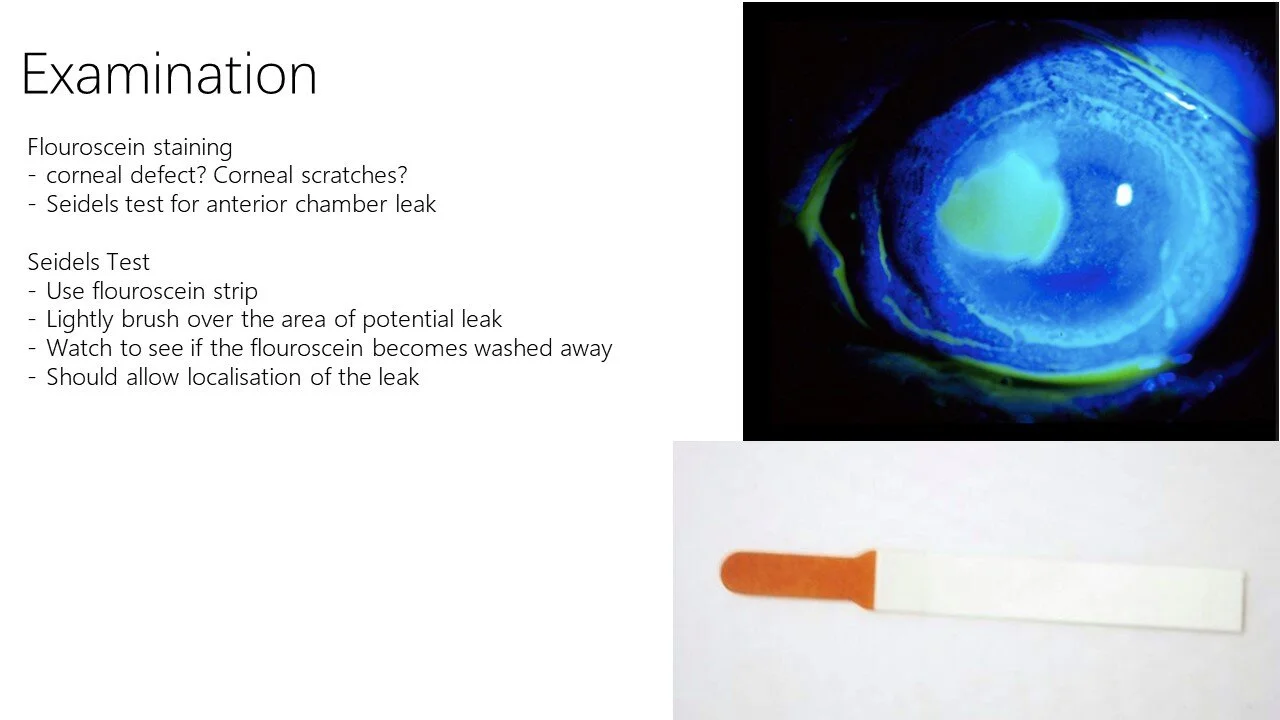
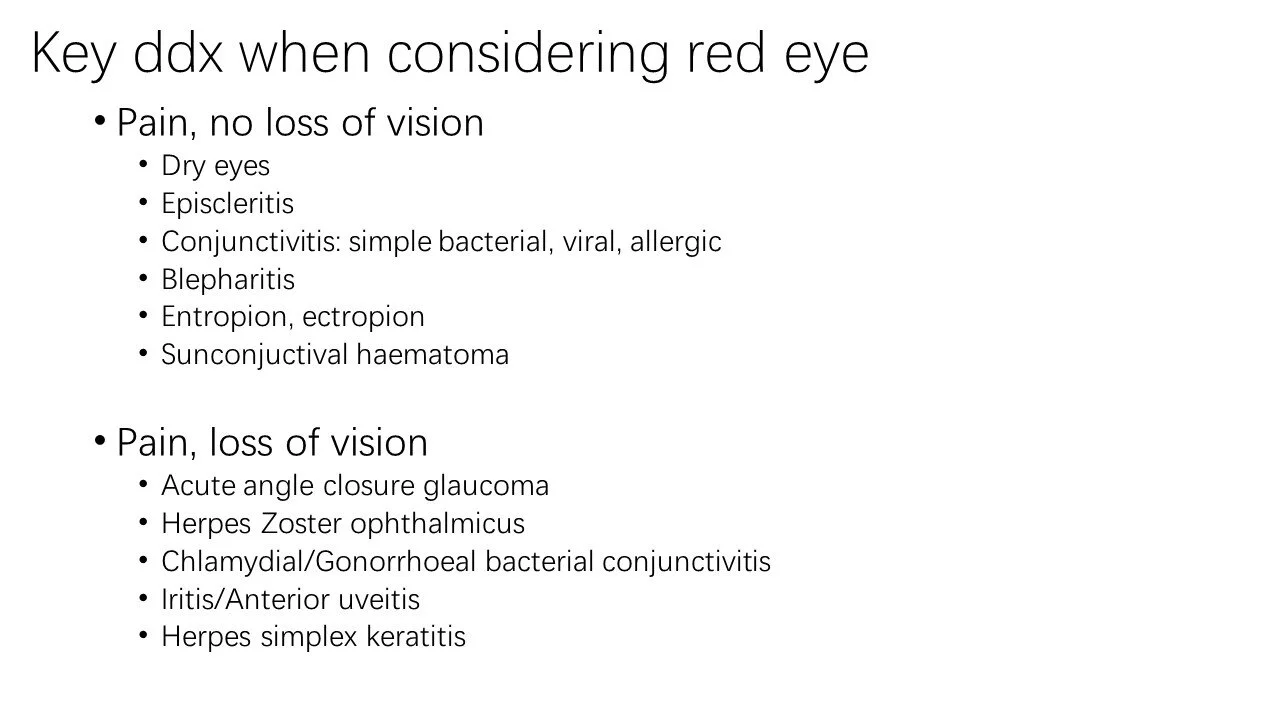
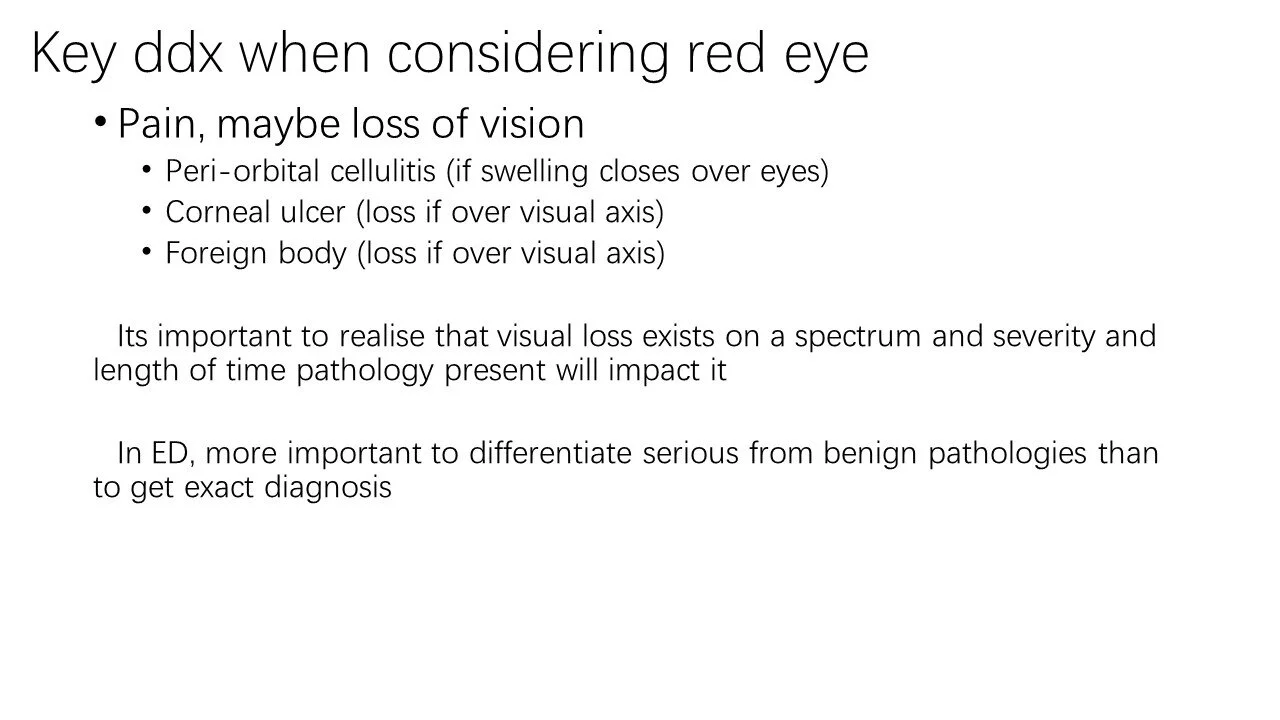
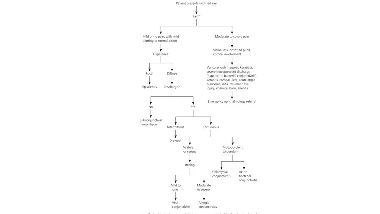
1130—1200: Differentials of the Red Eye — Luke Bayley
1200—1230: Causes of Diplopia — Andrew West
1230—1240: Learning from London’s 2020 COVID-19 Retrospectoscope — Sarah Simons
1240—1315: Lunch
1315—1415: Fellowship Written Group with Alex Handrinos (Zoom Meeting link) — Rachel Goh, Andrew West, Roshetha Bansal, Matthew Kilmurray
1345—1445: ALS Practical with Stuart Dilley (ED Handover Room) — Ezra Limm, Joseph Colcough, Alex Burman
1345—1445: Ultrasound with Mark Rugless — Rex Gavan
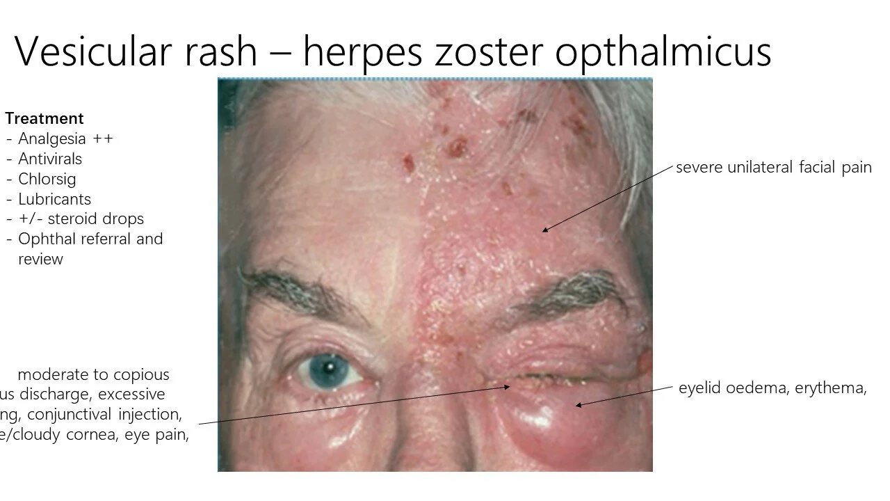
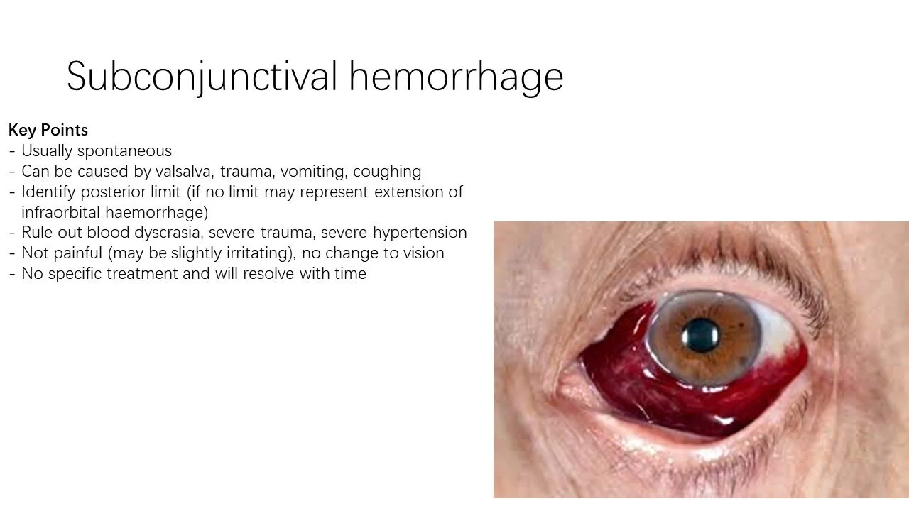
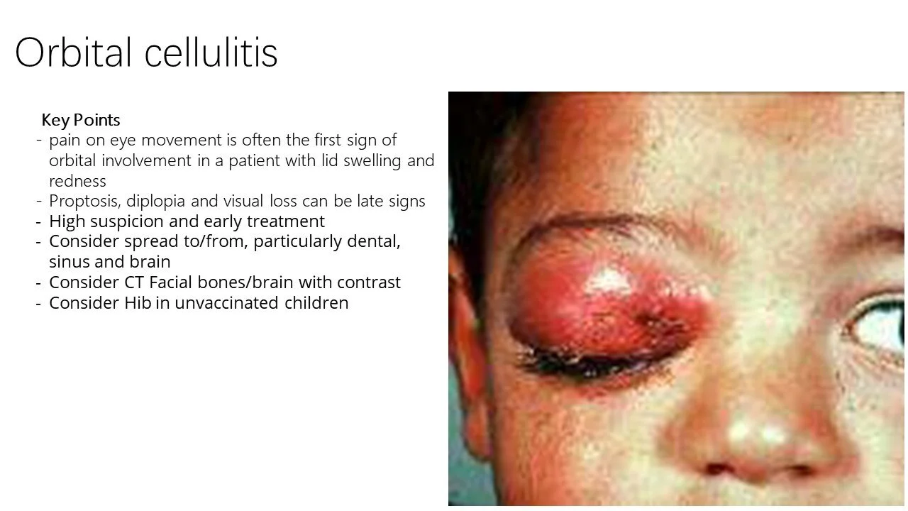
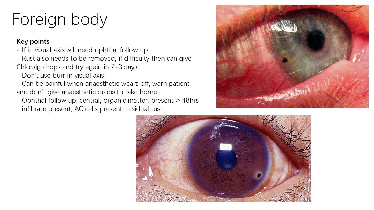
Resources
The Royal Victorian Eye & Ear Hospital has Clinical Practice Guidelines:
https://www.eyeandear.org.au/page/Health_Professionals/Knowledge_Portal/Eye_and_Ear_Emergency_Department/CPGs/
EYE AM GROOT
Acronym for causes of diplopia
Eye - Monocular causes — e.g. cataract
A - Aneurysm (must be excluded in the context of painful diplopia)
M - Myasthenia gravis
G - Gaze palsy — supranuclear, nuclear or internuclear
R - Reduced eye movement, i.e. ocular muscle entrapment — e.g. orbital floor fracture, cellulitis, retro-bulbar haematoma
O - Occipital lesion — e.g. mass, haemorrhage
O - Ocular nerve palsy (CN III, IV, VI)
T - Thyroid eye disease (Grave’s disease / Hyperthyroidism)
Therefore tests for diplopia:
CTB +/- Angio
MRI Brain — for gaze and ocular nerve palsies
TSH
Acetylcholine receptor antibodies (myasthenia gravis)
ECG of the Week
40yo lady presents drowsy.
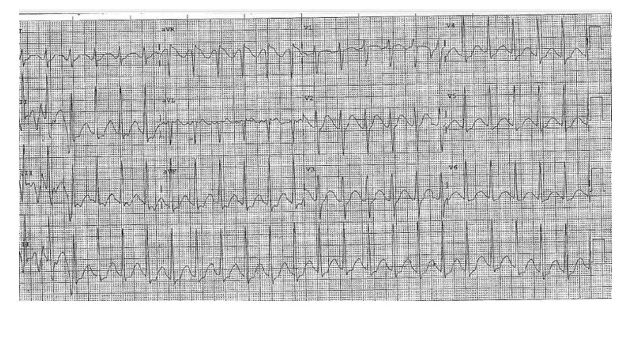
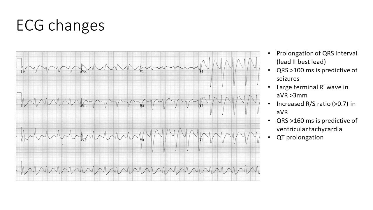
1) Describe the ECG
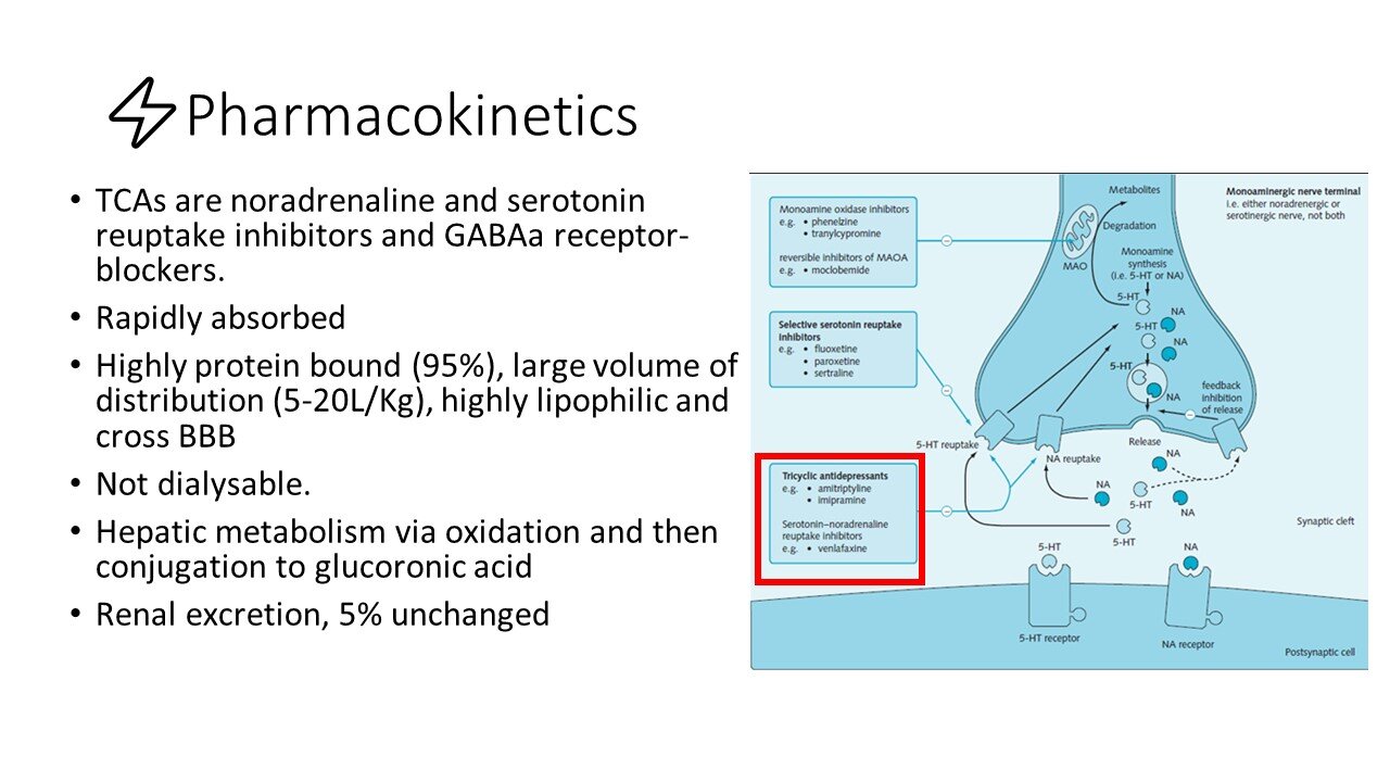
This patient took a dothiepin overdose (tricyclic antidepressant).
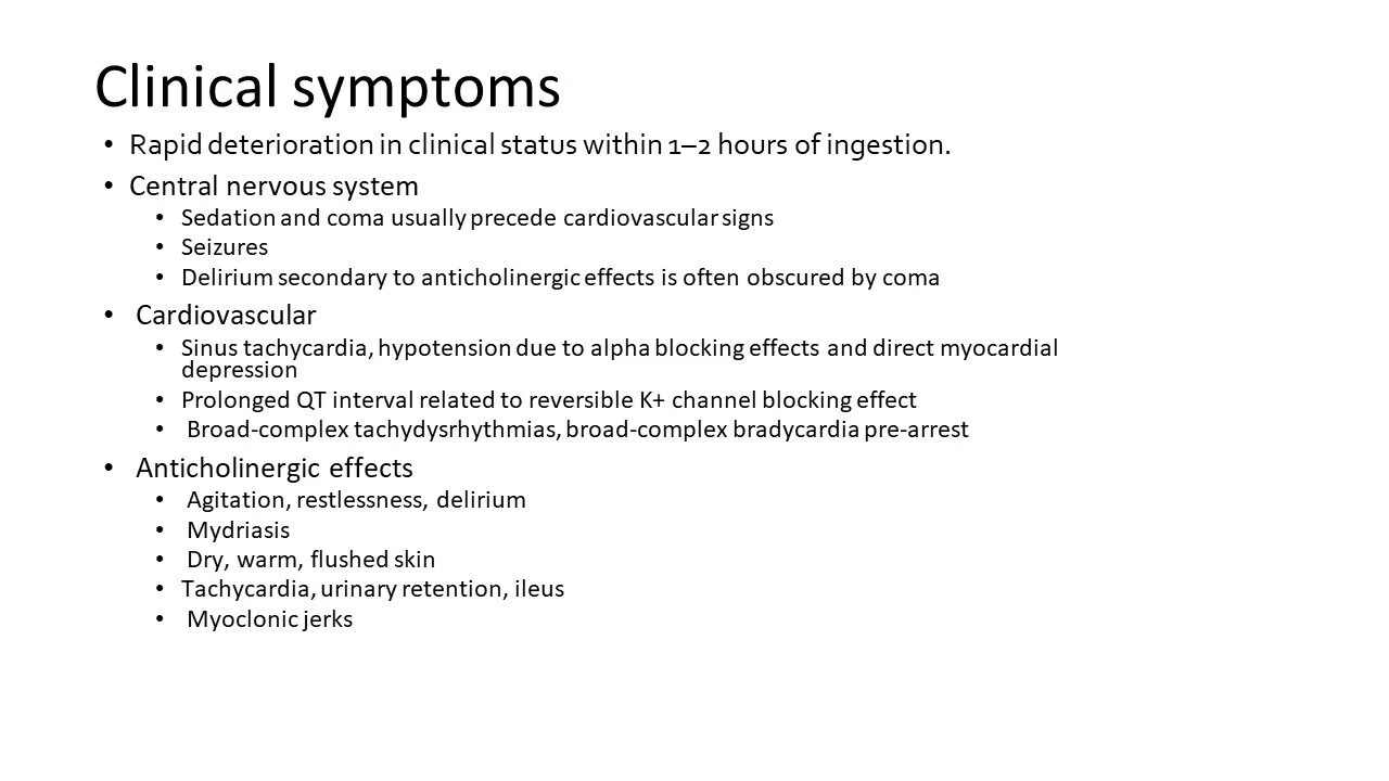
Sinus tachycardia (muscarinic-blocking effect of TCA)
p waves present but difficult to see — most obvious in aVL
Long QTc 500ms (potassium-blocking effect of TCA)
QRS duration 90-100ms (sodium-blocking effect of TCA)
Tall terminal R wave in aVR, not quite reaching the >3mm typical of TCA overdose (sodium-blocking effect of TCA)
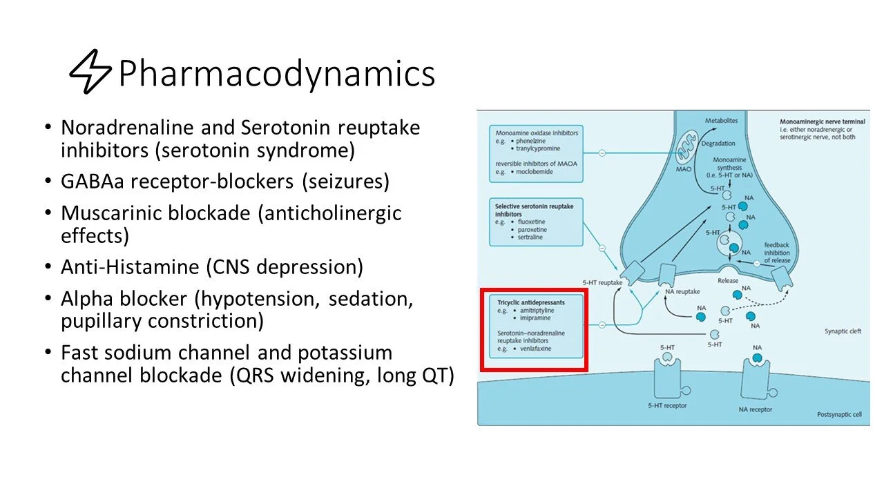
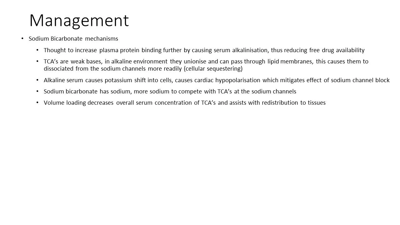
TCAs have a serotoninergic effect (block reuptake of serotonin) and also block potassium, sodium, calcium, muscarinic, peripheral alpha, histamine and GABA receptors.
Signs of toxicity on ECG:
Prolonged QRS (Na channel blockade) — this should be calculated from Lead II.
QRS >100ms = Predictive of seizures
QRS >160ms = Predictive of ventricular arrhythmias
Long QT (K channel blockade)
Tall terminal R wave in aVR >3mm (Na channel blockade)
R/S ratio >0.7 in aVR (Na channel blockade)
2) What agents have likely caused these changes? List at least 5.
Drugs that block Na channels and/or prolong the QT. For example:
TCAs
Anti-arrhythmics with Na channel blocking effects — flecainide, procainamide, propranolol
Antimalarial — hydroxychloroquine
Local anaesthetic — bupivicaine, ropivicaine
Carbamazepine
3) What is your general management plan?
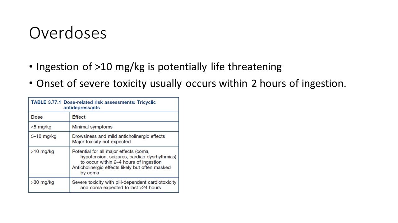
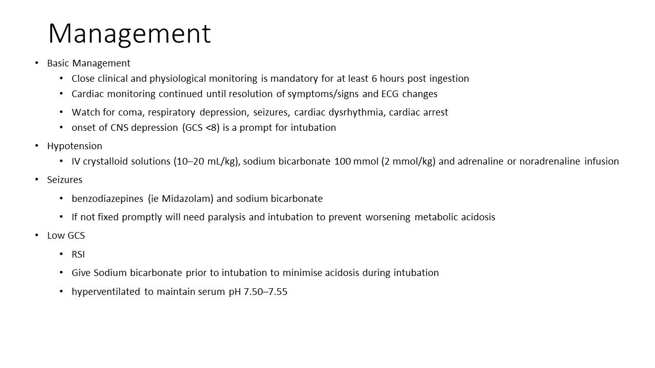
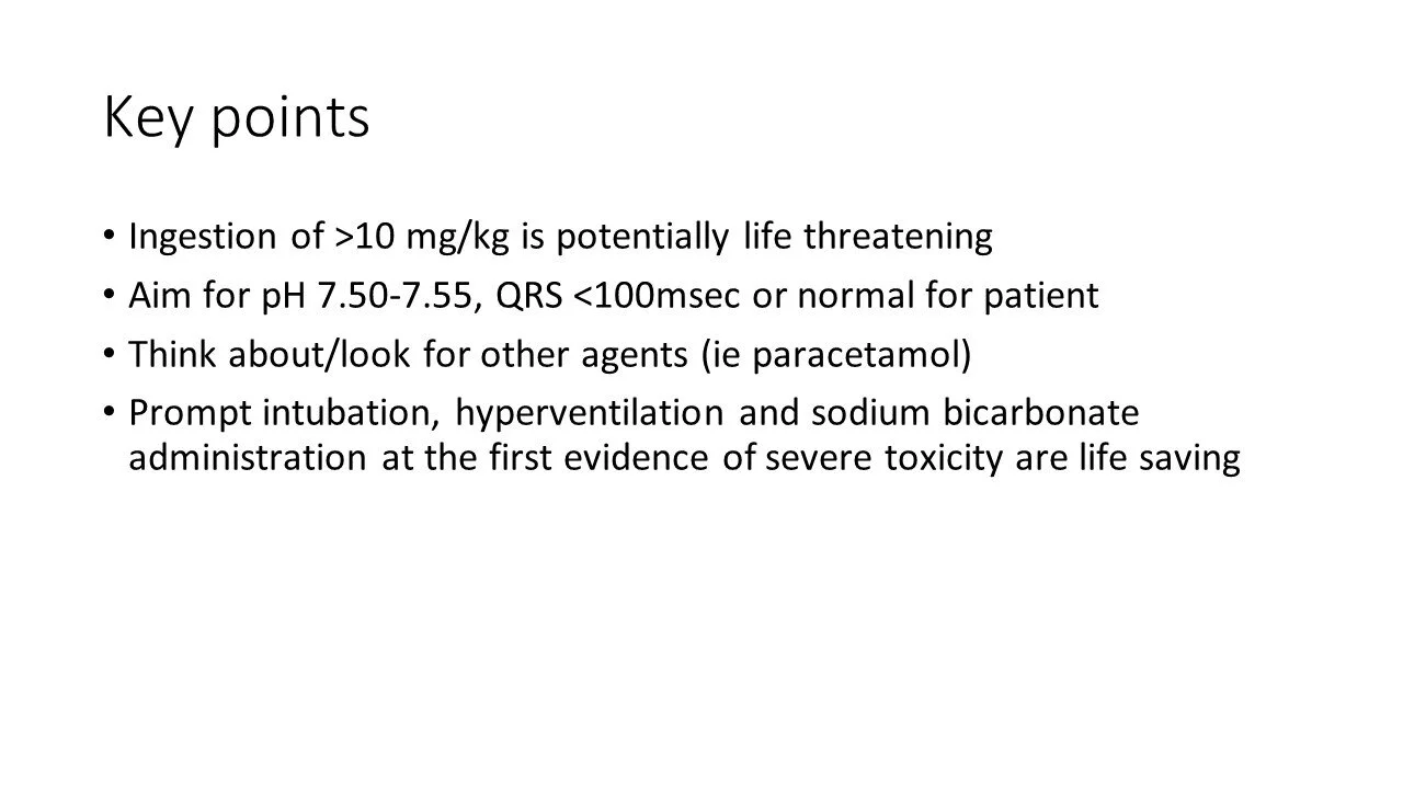
Determine risk — >10mg/kg is potentially life threatening. Severe toxicity usually manifests within 2 hours.
Start in Resus
Fluid resus — IV crystalloid 20mL/kg, repeat aiming for MAP 65
Low threshold for intubation (GCS <12) given tendency to rapidly deteriorate
Give IV sodium bicarb 8.4% 100mL prior to intubation to reduce risk of peri-intubation respiratory acidosis
Intubation also allows you to administer activated charcoal 50g
Once intubated, hyperventilate to pH >5.5
Minimum of 6 hours observation post-ingestion (cardiac monitoring until ECG changes resolve)
4) What specific treatments can be given?
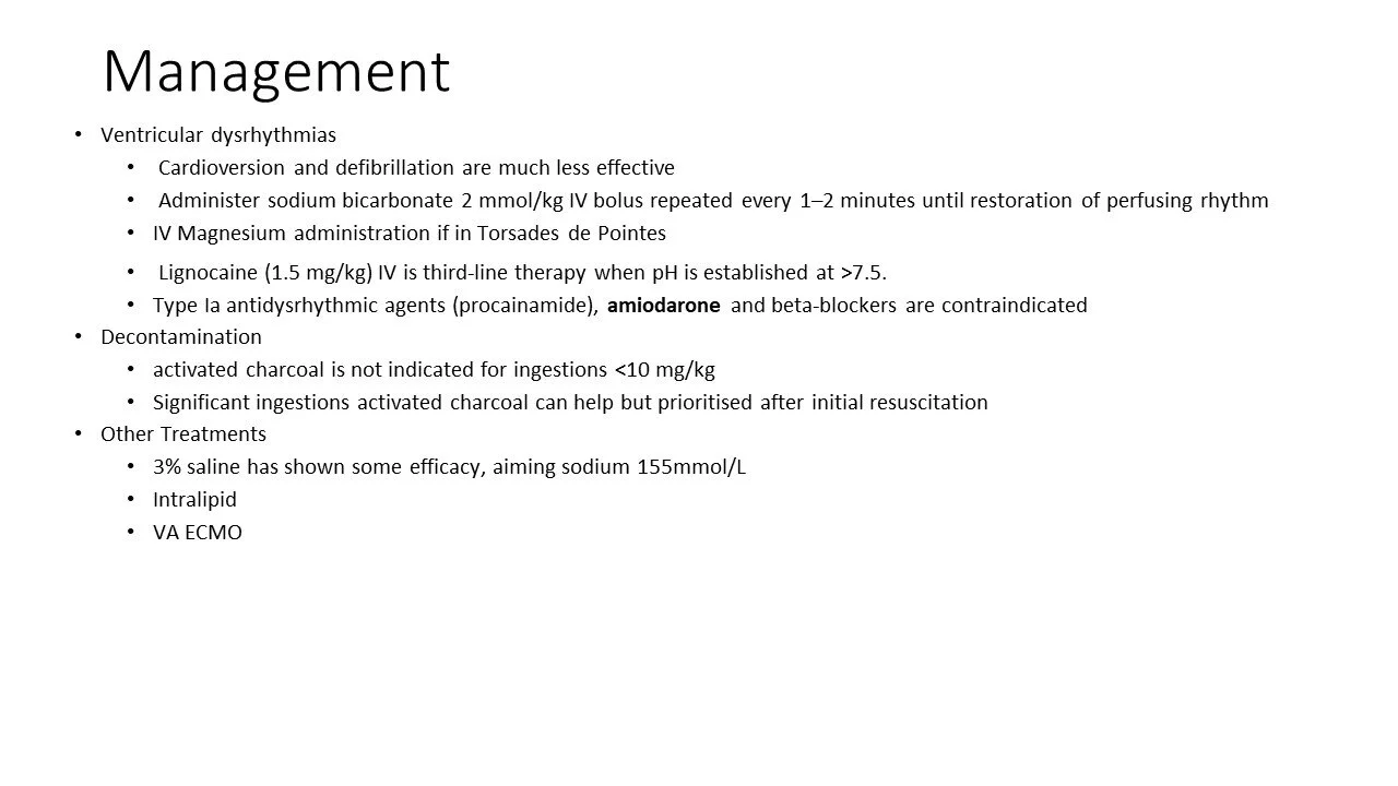
Treat cardiotoxicity / arrhythmias — IV sodium bicarb 8.4% 100mL, repeat every 5 minutes, aiming for resolution of hypotension, normal QRS and pH 5.5
Beware severe hypernatraemia from excessive NaHCO3
It’s the change in pH that matters (from acidosis to alkalosis), not so much the actual pH
Treat seizures — IV midazolam 1-2mg
Avoid flecainide, procainamide, anti-arrhythmics, beta-blockers and amiodarone as they may worsen hypotension and conduction abnormalities
Image of the Week
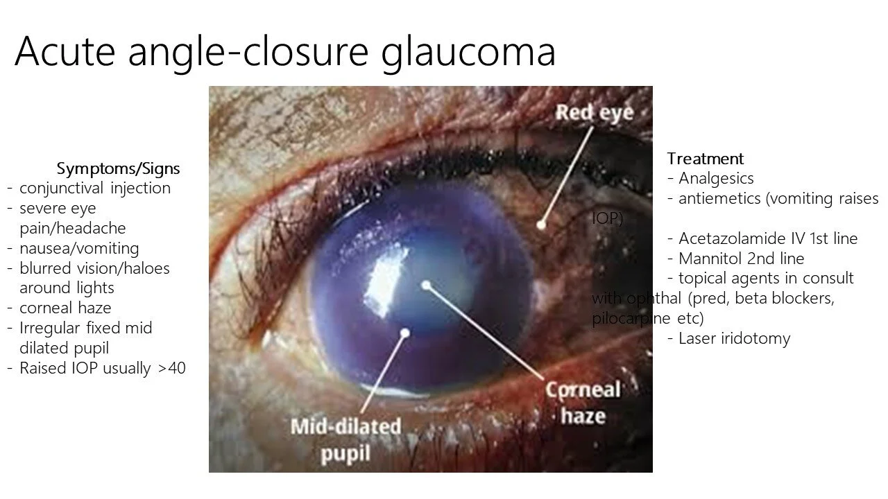
65yo lady presents with a painful red right eye.
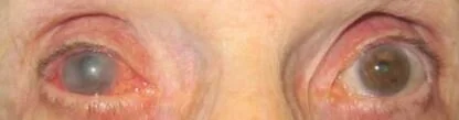
Image of the Week
65yo lady presents with a painful red right eye.
1) Based on the image, what is the most likely diagnosis and why?
Acute angle closure glaucoma
Mid-dilated pupil
Corneal oedema
Conjunctival injection
Elderly
2) Name 3 differential diagnoses
From front to back of eye:
Conjunctivitis
Keratitis
Corneal ulcer
Iritis
Scleritis
Trauma, e.g. chemical injury
Endophthalmitis
3) What do you expect the intraocular pressure to be?
Usually >35mmHg
4) Give 3 initial treatment options (include doses)
IV acetazolamide 500mg stat, then PO 250mg QID (reduces aqueous production)
Timolol 0.5% / Brimonidine 0.2% x 1 drop in affected eye every 30 minutes (reduces aqueous production)
Pilocarpine 2% x 1 drop in affected eye every 15 minutes (increases aqueous outflow by opening angle through miosis) — start after IOP < 40
IV mannitol 1g/kg (decreases vitreous volume) — if acetazolamide fails