12 May 2021 - Nervous System Disorders
Teaching this week will be over Zoom:
https://zoom.us/j/5981463478?pwd=RThaZzNkL2w5bldBMlNHeE1zZFFaZz09
Meeting ID: 598 146 3478
Passcode: 2021
Program
1000-1100: “Ask the Expert” — Neurologist Dr Lauren Sanders
1100-1130: Case Presentation — Laura Burns
NB: 1100-1130 Break-out group for those sitting Fellowship Written Exams (Andrew West, Jason Aw, Jonathan Leslie, Rachel Goh) with Alex Handrinos
1130-1140: Recap of Gastrointestinal and PEM Regional Teaching
1140-1150: Image of the Week — Mim Scharkie
1150-1200: ECG of the Week — Ben Sinnett
1200-1230: Lunch
1230-1400: M&M and S&S — Kirsten Cassidy
1400-1430: Registrar Forum — Mim Scharkie
Stroke Talk Recap
Here are some summary points from the stroke Q&A:
Timing of stroke therapies
When patient’s wake up with stroke symptoms, the “time of onset” becomes the midway point between when they went to sleep and when they woke up, e.g. if they slept from midnight to 8am, the “time of onset” is said to be 4am
On a case-by-case basis, some stroke patients might be thrombolysed beyond the 4.5 hours since onset of symptoms
Clot retrieval might be performed up to 24 hours after onset of symptoms if there is salvageable cerebral tissue on perfusion imaging
We should therefore call a Code Stroke and organise CTB/CTA and CT Perfusion on suspected stroke patients up to 24 hours post-symptom onset
The subtle neurological symptom
Strokes generally present with a neurological deficit rather than a positive finding
When the patient comes in complaining of tingling limbs, it needs to be distinguished whether this is a deficit (i.e. paraesthesia) or a positive finding
We can be reassured that patients with vague neurological symptoms will almost certainly not be candidates for thrombolysis or clot retrieval, and therefore time is not as important
Dizzy patient
These are hard patients to manage and stroke neurologists can find it difficult to distinguish what is and what isn’t a stroke
It is rare for a patient to present with dizziness due to a stroke without any other neurological findings. Thorough history and examination are key
Further resources
This free, 60 minute podcast on stroke management is a must listen to for all ED clinicians
Here is a great summary of the complex literature on thrombolysis, from an ED perspective
ACEM also have a statement on thrombolysis that should be read by all
Image of the Week
87yo lady tripped on grass, injuring her right eye. Past history of Parkinson’s (not on treatment).
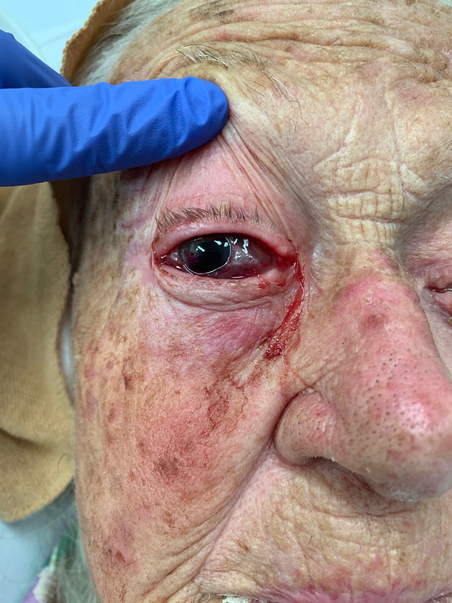
Describe the image.
Anterior chamber hyphaema (Grade 4)
Extravasation of anterior chamber
Clues there is a globe rupture (although you can’t actually tell this from this picture
Irregular shaped pupil
Enophthalmos (see image below)
Oedematous subconjunctival haemorrhage
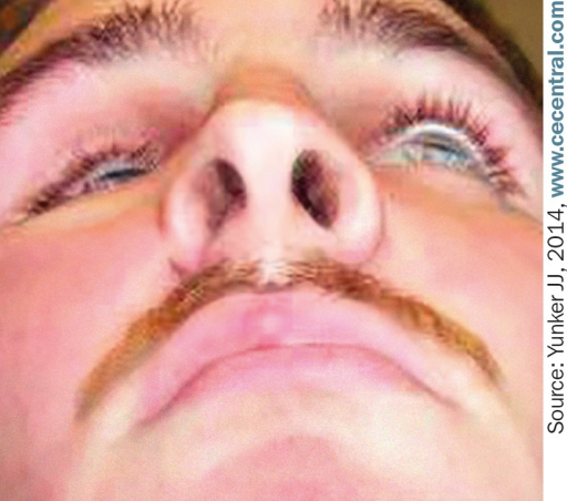
Enophthalmos of the right eye
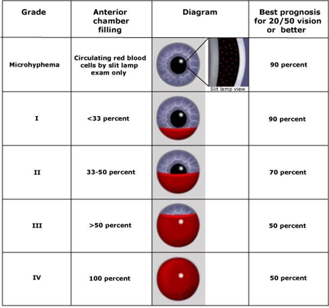
What is the classification for this injury?
Grade 1: Layered blood occupying less than one third of the anterior chamber
Grade 2: Blood filling one third to one half of the anterior chamber
Grade 3: Layered blood filling one half to less than total of the anterior chamber
Grade 4: Total clotted blood, often referred to as blackball or 8-ball hyphaema
How would you manage this injury?
Topical cyclopentolate 1% eye drop TDS to immobilise the rise and potentially stabilise the clot
Topical prednisolone 1%/phenylephrine 0.12% (Prednefrin Forte) eye drop QID to reduce trauma-associated inflammation
Quiet ambulation at home for 1-2 weeks (most re-bleeds occur within 3-5 days)
Sleep at 30 degrees (encourages hyphaema to settle) with an eye shield on (prevent further injury)
Avoid NSAIDs, and review indication for any anticoagulants that patient might be on
Ophthalmology review in 24-72 hours, watching for spike in intraocular pressure (due to blood blocking drainage of aqueous from the anterior chamber)
What are common complications with this injury?
Re-bleed (within 3-5 days)
Traumatic iritis
Vision loss
Infection
ECG of the Week
57yo man brought in by ambulance from a nightclub pool party, GCS 3 and wearing only a towel.
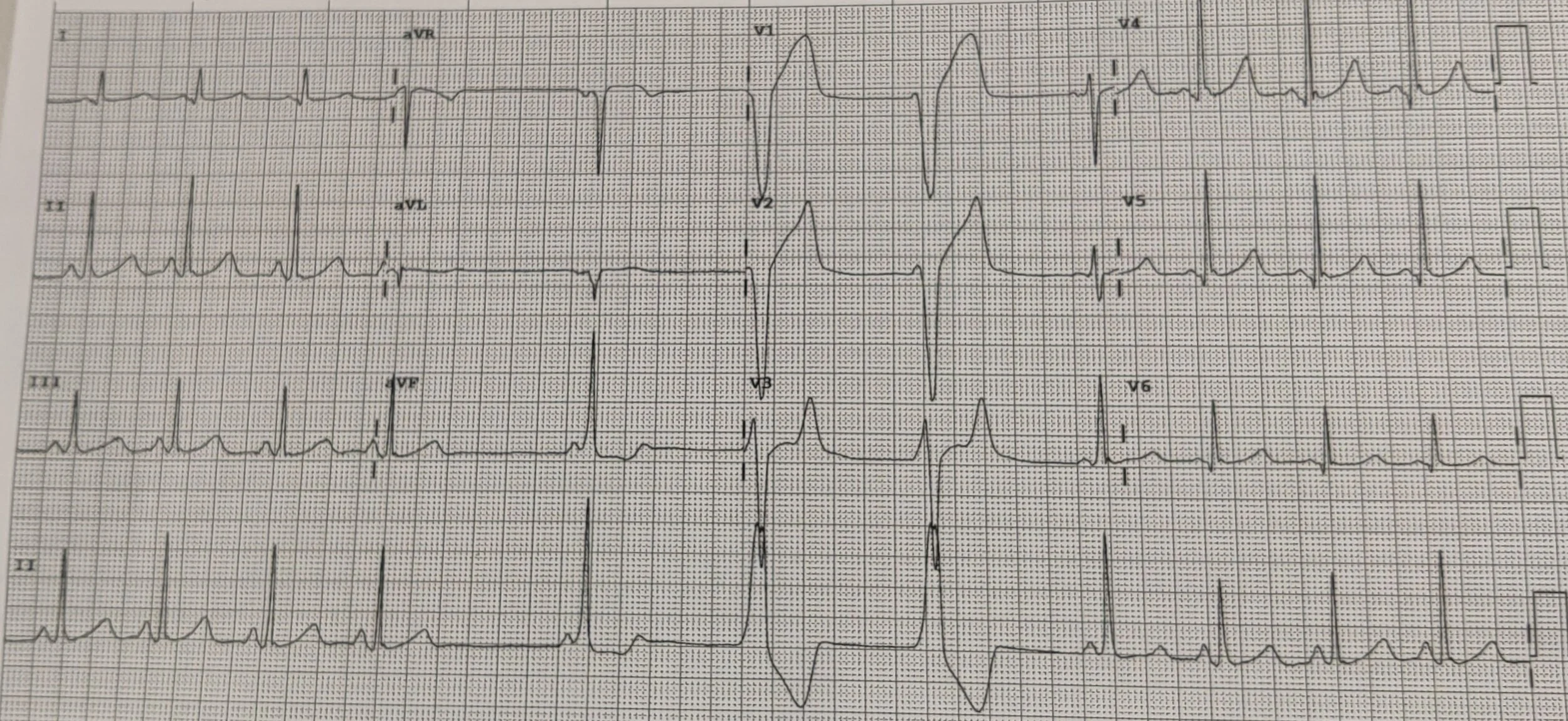
Describe the ECG.
Sinus rhythm HR 80bpm, normal axis, normal PR and QT intervals — until period of non-sustained bradycardia with atrial ectopic, followed by ventricular couplets. Small Osborn waves, most pronounced in precordial leads.
What is the most likely cause? Differentials?
In context of the stem + Osborn waves (also known as J waves) = hypothermia (patient's core temp was 31.5°C)
Other differentials = any that would account for myocardial irritability + altered mental status, e.g. electrolyte abnormality, intracranial event, tox, etc.
Based on your primary diagnosis, what other ECG findings might you see with this condition?
Sinus bradycardia (a normal physiological response, due to slowed cardiac depolarisation)
Slow AF / junctional bradycardia / AV block
QRS widening / QT prolongation [everything on the ECG gets slower / wider]
Shivering artefact
ST elevation (non-territorial; due to aberrant depolarisation)
The patient becomes hypotensive. Discuss how you'd manage this patient, including any targeted treatments.
If bradycardic, atropine is unlikely to be effective (not vagally mediated). Worth attempting, but efforts to warm the patient are key.
“They're not dead until they're warm and dead” (above 32°C)
Tympanic temperature is unreliable at < 34°C. Use a core temperature probe (e.g. oesophageal or bladder). Available from theatre if not in ED.
Circulating drug times will be increased = risk of toxic accumulation as the patient is warmed
Warm the patient
Remove wet clothing
Warming blankets / Bair hugger
Humidified O2
For the severely compromised, intrathoracic lavage (2 x large ICC, circulating heated fluids), dialysis or ECMO can be used
Give fluids (to counter the physiological hypothermic diuresis)
Give warmed fluids (to prevent iatrogenic cooling through crystalloid administration, rather than strictly warming the patient)
Monitor BSL (catecholamine stress response = initial hyperglycaemia > transient insulin resistance > risk of post-warming hypoglycaemia